IMAGE

Fig. 3

ID
ZDB-IMAGE-201121-33
Source
Figures for Kondrychyn et al., 2020
Image
Figure Caption

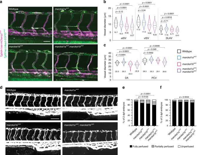
Fig. 3 Depletion of <italic>Marcksl1</italic> expression alters vessel diameter and delays lumenisation.

a Maximum intensity projection of confocal z-stacks of zebrafish trunk vessels of 2 dpf wildtype, marcksl1ark23, marcksl1brk24 and marcksl1ark23;marcksl1brk24 embryos in Tg(fli1ep:Lifeact-EGFP)zf495;Tg(kdr-l:ras-mCherry)s916 background. b, c Quantification of aISV, vISV and DLAV (b) and DA and PCV (c) in wildtype and marcksl1 mutant embryos (wildtype: n=26 aISVs/27 vISVs from 15 embryos; marcksl1ark23: n = 43 aISVs/63 vISVs from 29 embryos; marcksl1brk24: n = 79 aISVs/57 vISVs from 39 embryos; marcksl1ark23;marcksl1brk24: n = 74 aISVs/46 vISVs from 28 embryos). Data are collected from 2 (marcksl1ark23;marcksl1brk24 embryos), 3 (marcksl1ark23 and marcksl1brk24 embryos) and 4 (wildtype embryos) independent experiments. Violin plots represent the entire range of values, dotted lines indicate first and third quartiles, center lines are median. Mean values are indicated. Statistical significance was determined by ordinary one-way ANOVA with Tukey’s multiple comparisons test. df Microangiography in wild type and marcksl1 mutant embryos. Images are representative of wildtype, marcksl1ark23, marcksl1brk24 and marcksl1ark23;marcksl1brk24 embryos at 2 dpf. Quantification of lumenisation of ISVs in wildtype and marcksl1 mutant embryos at 2 dpf (e, n = 1472 ISVs from 32 wildtype embryos; n=1058 ISVs from 23 marcksl1ark23 embryos; n = 1150 ISVs from 25 marcksl1brk24 embryos; n=1564 ISVs from 34 marcksl1ark23;marcksl1brk24 embryos) and at 3 dpf (f, n = 1518 ISVs from 33 wildtype embryos; n = 1104 ISVs from 24 marcksl1ark23 embryos; n = 782 ISVs from 17 marcksl1brk24 embryos; n = 1748 ISVs from 38 marcksl1ark23;marcksl1brk24 embryos). ISVs no. 4-26 were analyzed on both sides of the embryo. Data are collected from three independent experiments and analyzed by ordinary one-way ANOVA with Dunnett’s multiple comparisons test. DA dorsal aorta; DLAV, dorsal longitudinal anastomotic vessel; ISV intersegmental vessel; aISV arterial ISV; vISV venous ISV; PCV posterior cardinal vein. Scale bars, 50 µm (a) and 100 µm (d). Source data are provided as a Source data file.

Figure Data
Acknowledgments
This image is the copyrighted work of the attributed author or publisher, and ZFIN has permission only to display this image to its users. Additional permissions should be obtained from the applicable author or publisher of the image. Full text @ Nat. Commun.