
|
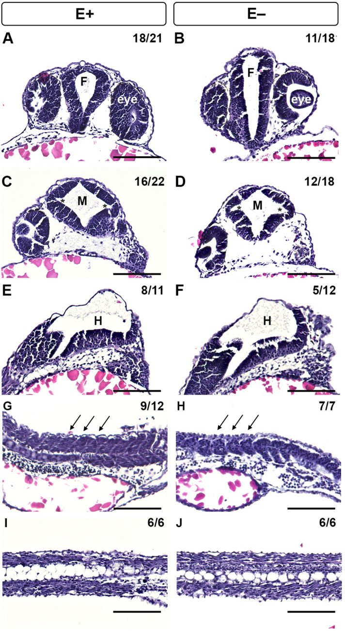
Figure 8
Histological analysis of 24 hpf zebrafish embryos with morphological defects associated with VitE status. Hematoxylin and eosin staining of E+ and E− embryos at 24 hpf was used to evaluate morphological defects, including fore- (F) and mid- (M), and hind- (H) brain ventricle inflation, somite formation and notochord vacuolation. Transverse section of E+ embryos had tear-drop shaped-F with eyes to each side (