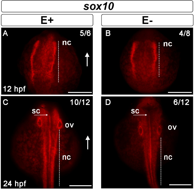
Neural crest cell migration impaired during development by VitE deficiency. Sox10 was expressed in the cells of the neural border at 12 hpf (A, B); arrows indicate dorsal region of the embryo. The extent of the distribution of neural crest cells (nc) expressing sox10 was greater (n = 4/8) in E+ embryos (A) relative to E− embryos (n = 5/6) (B). Measurements in representative embryos were E+ 174 μm relative to E− 145 μm. NC migrate ventrally away from the spinal cord (sc) to differentiate into cranial, cardiac, enteric and sensory neurons and glia. At 24 hpf, sox10 expression around the otic vesicle (ov) was clearly defined in E+ embryos (C) and less well-defined in E− embryos (D). In E+ embryos (C), the migratory NC that flank the developing sc show increased sox10 expression (n = 10/12), while E− embryos (D) had fewer migratory NC with sox10 expression (n = 6/12). Nonetheless, similar spinal cord (s) sox10 expression was observed in E+ and E− embryos. Scale bar represents 100 μm; representative embryos are shown. Figure panels were generated with the BZ- × 700 microscope, processed with BZ-X Analyzer Software with image adjustments equally applied across time points in Adobe Photoshop v21.2.1.
This image is the copyrighted work of the attributed author or publisher, and
ZFIN has permission only to display this image to its users.
Additional permissions should be obtained from the applicable author or publisher of the image.
Full text @ Sci. Rep.
Your Input Welcome
Thank you for submitting comments. Your input has been emailed to ZFIN curators who may contact you if
additional information is required.
Oops. Something went wrong. Please try again later.