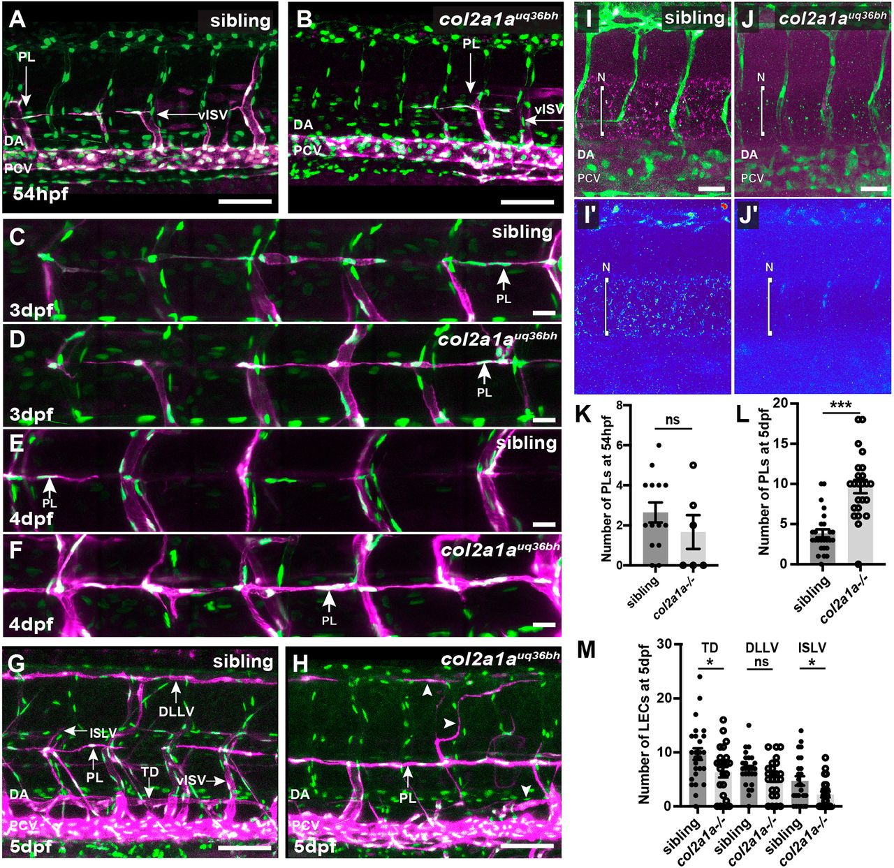
Fig. 3 col2a1auq36bh mutants exhibit increased PL numbers in the HM over time and reduced numbers of LECs in mature vessels. (A,B) Confocal images of Tg(fli1a:nEGFP); Tg(-5.2lyve1b:dsRed) sibling (A, n=16) and col2a1a1uq36bh (B, n=9) at 54?hpf showing PLs migrate to the HM. (C-F) Confocal images of Tg(fli1a:nEGFP); Tg(-5.2lyve1b:DsRed) sibling (C, n=4; E, n=7) and col2a1auq36bh (D, n=5; F, n=7;) embryos at 3?dpf (C,D) and 4?dpf (E,F) show that PLs fail to migrate from the HM in col2a1auq36bh mutants compared with the siblings. (G,H) Confocal images of sibling (G, n=32) and col2a1auq36bh (H, n=23) embryos at 5 dpf showing stalled PLs at the HM in col2a1auq36bh mutants. Arrowheads indicate vessel fragments. (I,J) IF assay of Col2a1 and eGFP in Tg(fli1a:nEGFP); Tg(-5.2lyve1b:dsRed) siblings (I, thermal map I?, n=24) and col2a1auq36bh (J, thermal map J?, n=8) mutants confirm loss of Col2a1a in col2a1auq36bh. (K,L) No difference in PL number was observed in col2a1auq36bh and siblings at 54?hpf (K); however, at 5 dpf there was a significant increase in stalled PLs at the HM in col2a1auq36bh mutants when compared with siblings (L). (M) Quantification of nuclei in the TD and ISLV revealed a significant reduction in col2a1auq36bh (n=23) when compared with siblings (n=32). Line marked N indicates notochord. ns, not significant. *P<0.05, ***P<0.001 (two-tailed unpaired Student's t-test). TD, thoracic duct; HM, horizontal myoseptum; ISLVs, intersegmental lymphatic vessels; N, notochord; PL, parachordal LEC. Scale bars: 50?Ám in A-H; 80?Ám in I,J.
Image
Figure Caption
Figure Data
Acknowledgments
This image is the copyrighted work of the attributed author or publisher, and
ZFIN has permission only to display this image to its users.
Additional permissions should be obtained from the applicable author or publisher of the image.
Full text @ Development

