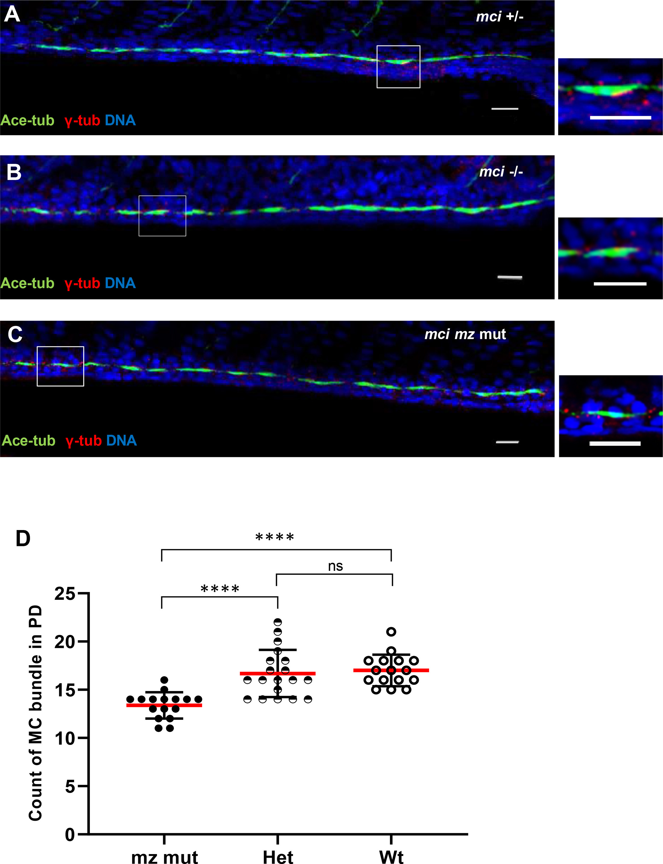
Fig. 5 Zebrafish embryos homozygous for a deletion allele of mci do not exhibit major defects in MCC formation within pronephric ducts. (A, B, C) Representative confocal images of pronephric ducts stained for cilia and basal bodies at 48 hpf. (A) mci heterozygote, (B) mci zygotic mutant, and (C) mci mz mutant. Scale bars = 20 μm. Highlighted areas (white box) are displayed as triplet of small panels. (D) Quantification of multicilia bundles in pronephric ducts of zebrafish embryos of different genotypes (mz mutant, heterozygous and wild-type for mci) Statistical analysis showed a significant decrease in number of these bundles in mz mutant embryos compared to heterozygote or wild-type embryos. Mean ± s.d., mean shown in red (N ≥ 16 embryos, ∗∗∗∗p < 0.0001, ns-not significant, Mann-Whitney U test).
Reprinted from Developmental Biology, 465(2), Zhou, F., Rayamajhi, D., Ravi, V., Narasimhan, V., Chong, Y.L., Lu, H., Venkatesh, B., Roy, S., Conservation as well as divergence in Mcidas function underlies the differentiation of multiciliated cells in vertebrates, 168-177, Copyright (2020) with permission from Elsevier. Full text @ Dev. Biol.

