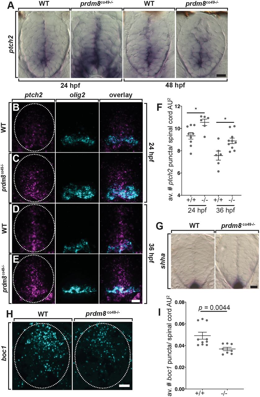
Fig. 9 Spinal cord cells of prdm8 mutant embryos have elevated Shh signaling activity. (A) Representative transverse sections of trunk spinal cords obtained from 24 and 48?hpf wild-type (WT) and prdm8co49?/? embryos (dorsal up) showing ptch2 RNA expression. (B-E) Representative transverse trunk spinal cord sections processed for fluorescent ISH to detect olig2 (blue) and ptch2 (pink) mRNA at 24?hpf (B,C) and 36?hpf (D,E). (F) prdm8co49?/? embryos have more ptch2 puncta per AU2 of spinal cord at 24?hpf (n=6) and 36?hpf (n=10) than wild-type embryos at 24?hpf (n=9) and 36?hpf (n=6). (G) Representative transverse sections of trunk spinal cord (dorsal up) showing shha RNA expression in 24?hpf wild-type and prdm8co49?/? embryos. (H) Representative transverse trunk spinal cord sections processed for fluorescent ISH to detect boc1 (blue) mRNA at 24?hpf. (I) Wild-type embryos (n=6) have more boc1 puncta per AU2 of spinal cord at 24?hpf than prdm8co49?/? embryos (n=8). Data are meanąs.e.m. with individual data points indicated. Statistical significance was evaluated by an unpaired, two-tailed Student's t-test (F) and by Mann-Whitney U test (I). *P<0.001. Dashed oval outlines the spinal cord boundary. Scale bars: 10??m.
Image
Figure Caption
Figure Data
Acknowledgments
This image is the copyrighted work of the attributed author or publisher, and
ZFIN has permission only to display this image to its users.
Additional permissions should be obtained from the applicable author or publisher of the image.
Full text @ Development

