IMAGE

Figure 3

ID
ZDB-IMAGE-200821-10
Genes
Source
Figures for Silic et al., 2020
Image
Figure Caption

Figure 3

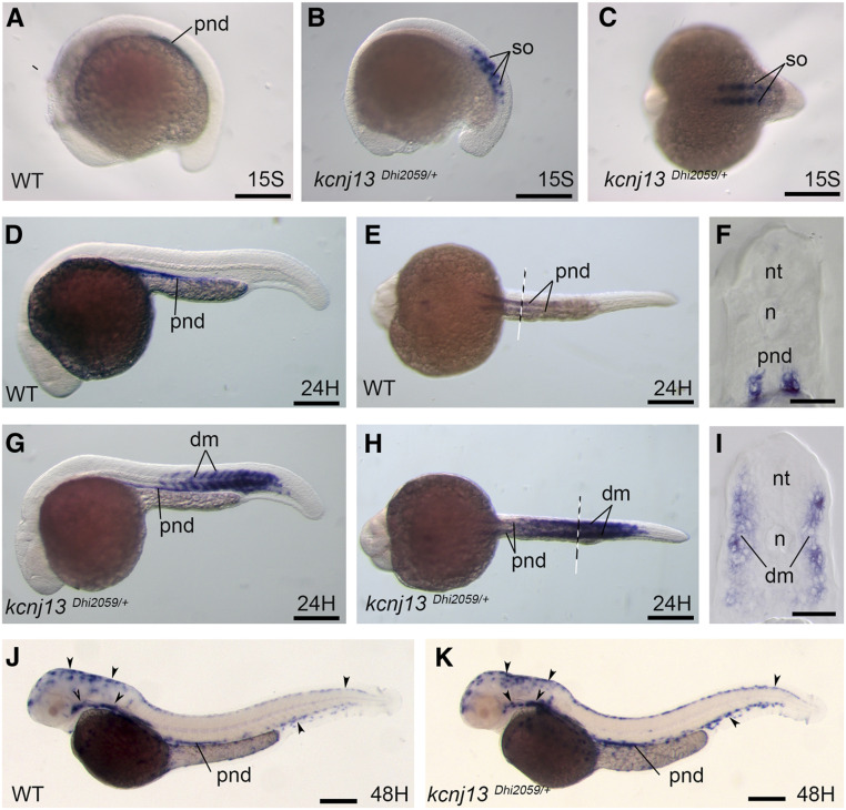
Kcnj13 is ectopically expressed in somites of the Dhi2059 mutant. Whole-mount in situ hybridizations of zebrafish embryos at the stages from 15S to 48 hpf were performed. WT embryos (A, D–F, and J). kcnj13Dhi2059/+ fish embryos (B, C, G–I, and K). (A) Kcnj13 is expressed at the pnd in WT embryos. (B) In the kcnj13Dhi2059/+ mutant, kcnj13 is expressed in the somites at the 15S stage. (C) Dorsal view of the embryo in (B). (D–F) At the 24-hpf stage, kcnj13 is continually expressed in the pronephric ducts of WT embryos. (D) Lateral view. (E) Ventral view of the 24-hpf embryos. The dashed line indicates the approximate position of the transverse section in (F). (G–I) The expression of kcnj13 in the kcnj13Dhi2059/+ mutant at the 24-hpf stage. The kcnj13 mRNA is also expressed in the somites in addition to pnds. (H) Ventral view of the same embryo in (G). The dashed line indicates the position of the transverse section in (I). (I) Transverse section revealed that kcnj13 is expressed in the dm. (J) At the 48-hpf stage, kcnj13 is continually expressed in pnds in WT fish embryos, and it is also expressed in melanocytes (arrowheads). (K) In the kcnj13Dhi2059/+ fish embryos, kcnj13 expression is similar to WT embryos at 48 hpf. The somite expression is no longer detectable. Bar, 250 μm (A–E, G, H, J, and K). Bar, 100 μm (F and I). 15S, 15-somite; dm, dermomyotome; hpf, hours postfertilization; mRNA, messenger RNA; n, notochord; nt, neural tube; pnd, pronephric ducts; so, somite, WT, wild-type.

Figure Data
Acknowledgments
This image is the copyrighted work of the attributed author or publisher, and ZFIN has permission only to display this image to its users. Additional permissions should be obtained from the applicable author or publisher of the image. Full text @ Genetics