
|
Fig. 7
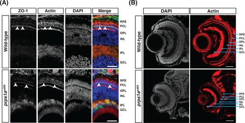
Cell polarity and retinal lamination are unaffected in prps1asd59 mutants. A, Representative maximum intensity projections of confocal images of transverse cryosections from wild?type and prps1asd59 mutant embryos at 5 dpf, stained for ZO?1 (green), actin (red), and 4′,6?diamidino?2?phenylindole (DAPI) (blue). No change in ZO?1 localization is observed in prps1asd59 mutants compared to wild?type. Arrowheads indicate ZO?1 localization to the apical membrane of the photoreceptor cell layer. GCL, ganglion cell layer; INL, inner nuclear layer; IPL, inner plexiform layer; OPL, outer plexiform layer; PCL, photoreceptor cell layer; RPE, retinal pigmented epithelium. Scale bars: 20 μm (n = 6 embryos for each genotype). B, Representative maximum intensity projections of confocal images of transverse cryosections from wild?type and prps1asd59 mutant embryos at 5 dpf, stained for actin and DAPI. Retinal patterning is unaffected in prps1asd59 mutants. Scale bars: 20 μm (n = 6 embryos for each genotype)