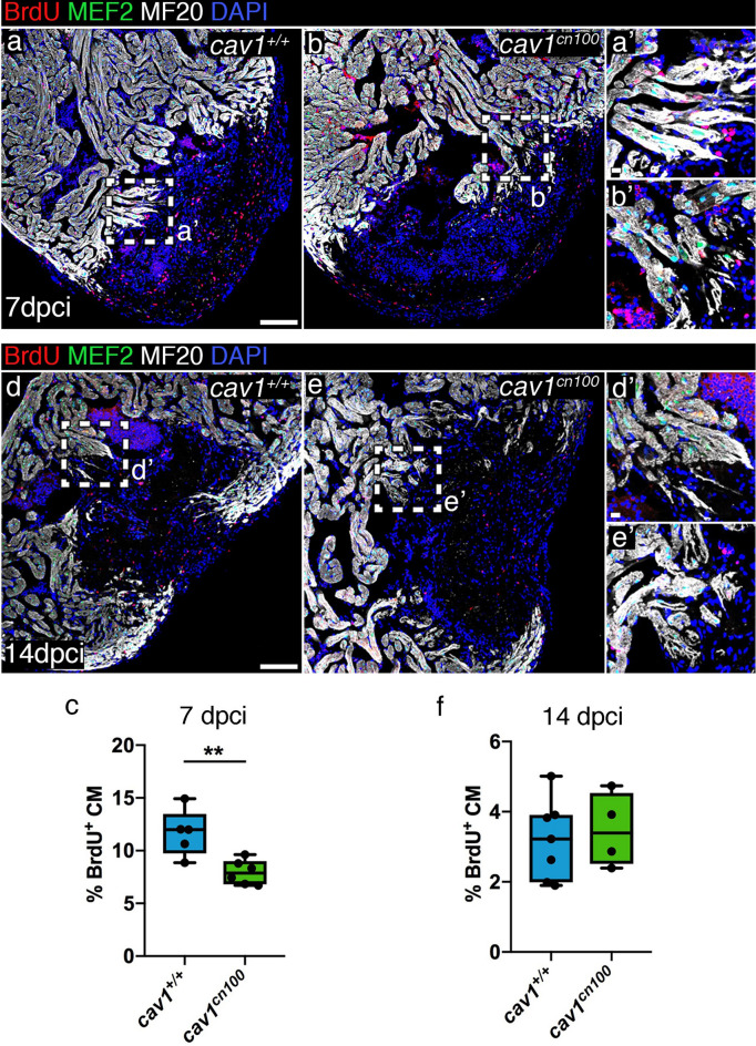
Cardiomyocyte proliferation is transiently reduced upon cryoinjury in cav1cn100 hearts. (a, b) Immunolabelling of 7 dpci cav1+/+ and cav1cn100 hearts for BrdU, MEF2 and MF20. (a′, b′) Magnifications of the dashed areas in (a) and (b). (c) Cardiomyocyte proliferation rate was measured by quantifying the BrdU+/MEF2+ nuclei to the total cardiomyocyte number in a 100 μm radius around the injured area. CM, cardiomyocytes. nWT = 5, ncn100 = 6, t-test, **P < 0.01. (d, e) Immunostaining of 14 dpci cav1+/+ and cav1cn100 hearts for BrdU, MEF2 and MF20. (d′, e′) Magnifications of the dashed areas in (d) and (e). (f) Quantification of cardiomyocyte proliferation at 14 dpci. nWT = 7, ncn100 = 4, t-test. Scale bars: 100 μm in (a), (b), (d), (e); 10 μm in (a′), (b′), (d′), (e′).
Acknowledgments
This image is the copyrighted work of the attributed author or publisher, and
ZFIN has permission only to display this image to its users.
Additional permissions should be obtained from the applicable author or publisher of the image.
Full text @ Sci. Rep.
Your Input Welcome
Thank you for submitting comments. Your input has been emailed to ZFIN curators who may contact you if
additional information is required.
Oops. Something went wrong. Please try again later.