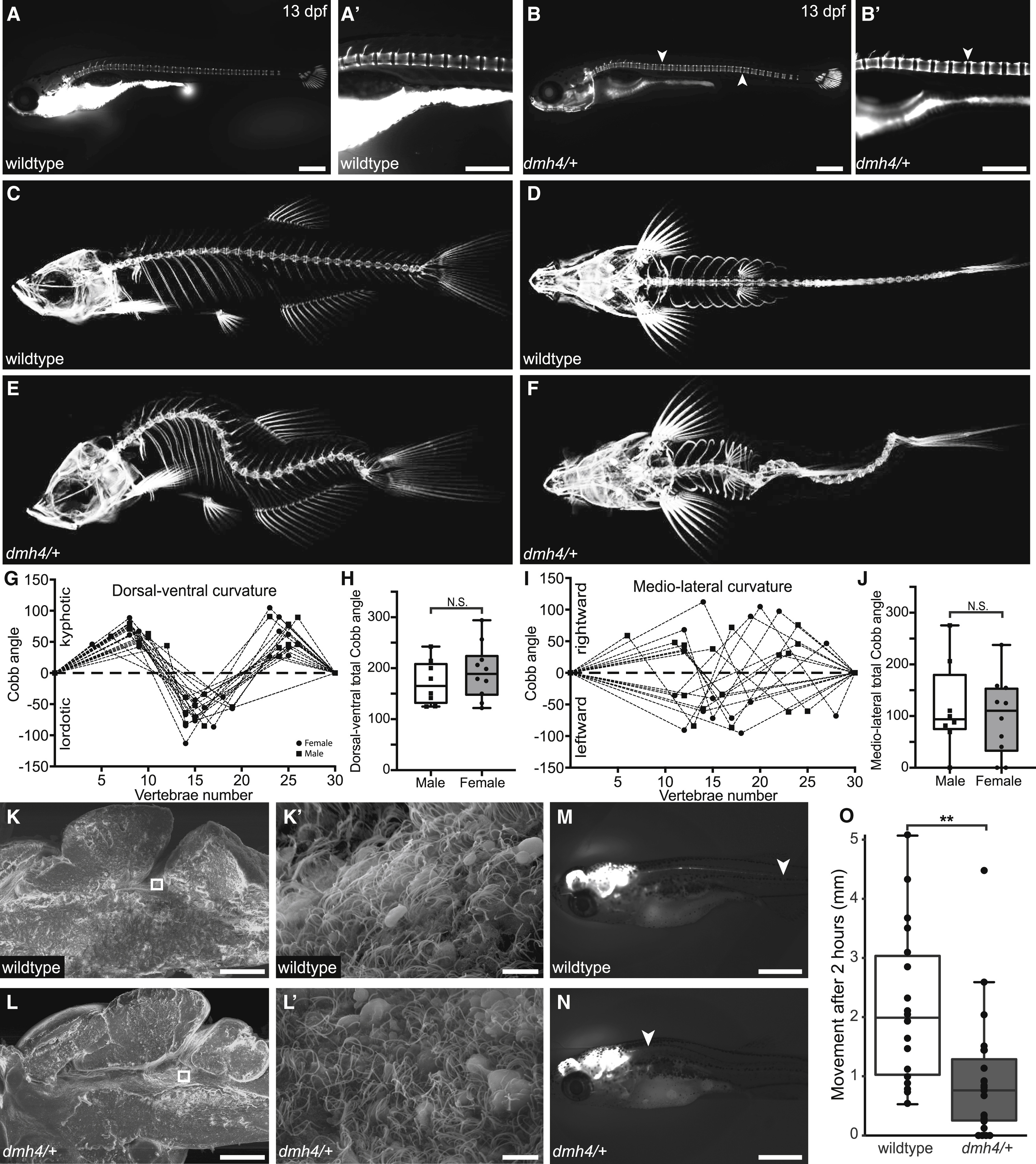
Fig. 1 Idiopathic-like Scoliosis in dmh4/+ Zebrafish Is Associated with Compromised CSF Flow in the Absence of Obvious Motile Cilia Defects (A and B) Calcein stains of wild-type (A and A?; N = 3; n = 24) and dmh4/+ (B and B?; N = 3; n = 29) larva at 13 dpf (6.5 mm standard length). Note the onset of spinal curvatures in dmh4/+ zebrafish (arrowheads) in the absence of congenital vertebral malformations. Scale bars represent 500 ?m. (C?F) Lateral (C and E) and dorsal (D and F) projections of three-dimensional microCTs for adult wild-type (C and D) and dmh4/+ (E and F) fish. (G?J) Quantification of curve severity, direction, and position along dorsal-ventral (G) and mediolateral (I) axes. Dashed lines indicate a Cobb angle of 0°, as per a wild-type spine. Data points for male and female fish are indicated as squares and circles, respectively. Combined total Cobb angle in dorsal-ventral (H; p = 0.4) and medio-lateral (J; p = 0.67) axes for males and females is shown. (K and L) Representative SEM images of wild-type (K; N = 3; n = 7) and dmh4/+ (L; N = 3; n = 10) bisected brains at 3 months of age. Scale bars represent 200 ?m. Squares indicate location of higher magnification images (K? and L?). Scale bars represent 5 ?m. (M?O) Representative images (M and N) and quantification (O) of bulk CSF movement along the spinal cord of 20-dpf wild-type (M; n = 18) and dmh4/+ (N; n = 18) larva, 2 h after injection of reporter dye into brain ventricles. p = 0.0038. Arrowheads indicate movement of dye. Scale bars represent 1 mm. See also Figure S1.
Image
Figure Caption
Figure Data
Acknowledgments
This image is the copyrighted work of the attributed author or publisher, and
ZFIN has permission only to display this image to its users.
Additional permissions should be obtained from the applicable author or publisher of the image.
Full text @ Curr. Biol.

