
|
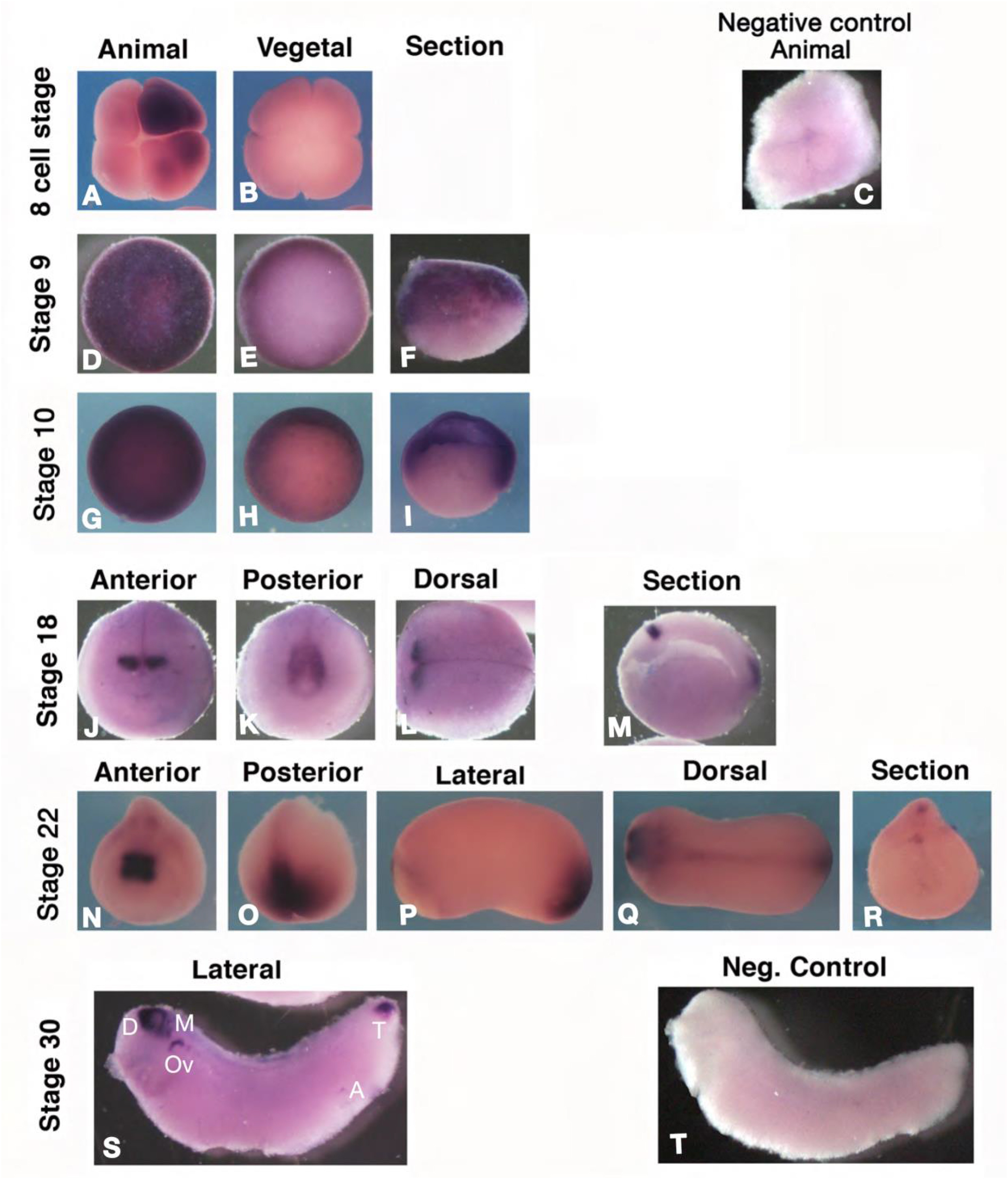
Fig. 1 Expression of Xenopus apcdd1. A-T. RNA in situ hybridization for Xenopus apcdd1. Maternal expression at 8 ?cell stage (A, B) is localized in the animal pole and is frequently unevenly distributed. In late blastulas (stage 9, D-F), the expression is animal and marginal (arrows in F, blastocoel in outlined in F). At the start of gastrulation (stage 10, G-I) apcdd1 RNA is present in animal and marginal cells, including the dorsal lip with the Spemann organizer (arrow in H, I, blastocoel is outlined in I). In neurula embryos (stage 18, K-N) and tailbud (stage 22), expression is limited to diencephalon and midbrain precursors (J, K, M, N, Q), and the tailbud (K, M, O, P, Q). Arrowhead in the transversal section (R) indicates ependymal precursors. In tadpoles (stage 30, S) expression closely mirrors that in mouse embryos. D ? diencephalon; M ? midbrain; Ov ? otic vesicle; T ? tailbud; A ? anus. Bracket in S indicates the branchial arches. For control in situ hybridization (C: 8-cell stage; T: tadpole), the RNA probe used was LacZ. The scale bar in A is 0.3 ?mm (all embryos are shown at the same scale). At least 10 Xenopus embryos were examined for each time point.
Reprinted from Developmental Biology, 464(1), Vonica, A., Bhat, N., Phan, K., Guo, J., Iancu, L., Weber, J.A., Karger, A., Cain, J.W., Wang, E.C.E., DeStefano, G.M., O'Donnell-Luria, A.H., Christiano, A.M., Riley, B., Butler, S.J., Luria, V., Apcdd1 is a dual BMP/Wnt inhibitor in the developing nervous system and skin, 71-87, Copyright (2020) with permission from Elsevier. Full text @ Dev. Biol.