IMAGE

Fig. 1

ID
ZDB-IMAGE-200619-30
Source
Figures for Loh et al., 2020
Image
Figure Caption

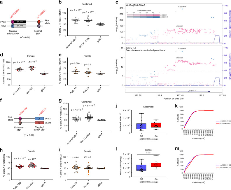
Fig. 1 Allelic expression imbalance of <italic>RSPO3</italic> and histological studies in abdominal and gluteal AT.

a Schematic illustration showing alleles of the sentinel SNP rs72959041 and the tagging SNP rs577721086. b Allelic expression analysis of RSPO3 transcripts was performed by qRT-PCR in abdominal and gluteal AT cDNA from 14 heterozygous carriers at rs72959041 (seven females, seven males). The proportion of total cDNA and gDNA containing the (WHR-increasing) rs577721086-C allele (r2 = 0.98 with rs72959041-A) is quantified on the y-axis. Error bars are median values with 95% confidence intervals. Statistical significance was assessed by two-tailed paired Student’s t-tests versus gDNA. cRSPO3 WHRadjBMI GWAS signals co-localise with subcutaneous adipose cis-eQTLs. d, e Allelic expression of RSPO3 transcripts were analysed in abdominal and gluteal isolated adipocyte (ADS) (d) and cultured AP (e) cDNAs from ten heterozygous female carriers at rs72959041 as in b. f Schematic illustration showing alleles of the enhancer SNP rs9491696 and the tagging SNP rs1892172. gi Allelic expression analysis of RSPO3 in g abdominal and gluteal AT cDNA from 32 individuals (15 females, 17 males) and in h abdominal and gluteal ADS and i cultured AP cDNA from 17 and 19 females, respectively, who are heterozygous at rs9491696. All individuals selected are homozygous for the rs72959041-G (non-risk) allele. The proportion of cDNA and gDNA containing the WHRadjBMI-increasing rs1892172-A allele (r2 = 0.84 with rs9491696-G) is quantified on the y-axis. Error bars are median values with 95% confidence intervals. Statistical significance was assessed by two-tailed paired Student’s t-tests versus gDNA. jm Median adipocyte cell weight calculated from median cell area measured from j abdominal and l gluteal AT histological sections from 14 and 15 pairs, respectively, of age- and BMI-matched females grouped by rs72959041 genotype. Cumulative frequency distribution of the adipocyte cell-surface area are shown for abdominal (k) and gluteal (m) AT. More than 250 cells were measured for each biopsy. Box and whisker plot: centre line, median; box limits, upper and lower quartiles; and whiskers, maximum and minimum values. Statistical significance was assessed by a two-tailed Wilcoxon signed-rank test. Source data are provided as a Source Data file.

Acknowledgments
This image is the copyrighted work of the attributed author or publisher, and ZFIN has permission only to display this image to its users. Additional permissions should be obtained from the applicable author or publisher of the image. Full text @ Nat. Commun.