Fig. 5
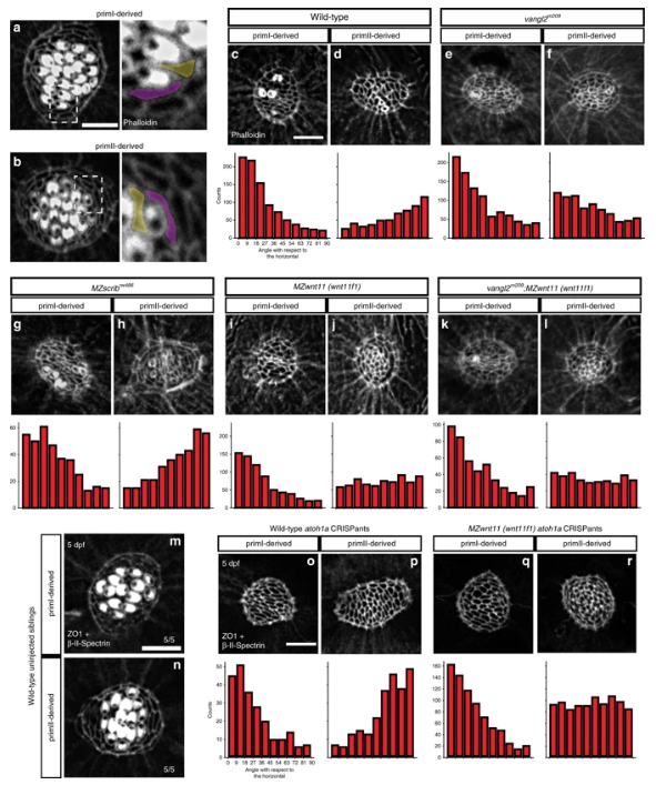
Wnt pathway genes are required for proper coordinated support cell organization. a, b Phalloidin staining of wild-type primI and primII-derived neuromasts. The dotted square outlines the area magnified in (a) and (b). Two support cells each were false colored to depict their orientation. b?j Phalloidin staining of neuromasts 3?h after hair cell ablation. c, d Wild-type primI (c) and primII-derived (d) neuromasts. e, f vangl2 mutant primI (e) and primII-derived (f) neuromasts. g, h MZscrib mutant primI (g) and primII-derived (h) neuromasts. i, j MZwnt11 (wnt11f1) mutant primI (i) and primII-derived (j) neuromasts. k, l vangl2;MZwnt11 (wnt11f1) double mutant primI (k) and primII-derived (l) neuromasts. The histograms show the distribution of binned angles of cell orientation with respect to the horizontal for each of the conditions tested. c, d Angle distribution in wild-type primI (c one-way chi square test p?=?1.78?×?10?87) and primII-derived (d p?=?4.54?×?10?19) neuromasts. Note that primI distribution is skewed toward being horizontally aligned, while the distribution in primII-derived neuromasts is toward vertical alignment. e, f Angle distribution in vangl2 mutant primI (e p?=?4.91?×?10?13) and primII-derived (f p?=?2.14?×?10?16) neuromasts. g, h Angle distribution in MZscrib mutant primI (g p?=?1.40?×?10?14) and primII-derived (h p?=?9.60?×?10?13) neuromasts. i, j Support cell orientation in MZwnt11 (wnt11f1) mutant primI (i p?=?5.32?×?10?52) and primII-derived (j p?=?0.006) neuromasts. k, l Support cell orientation in vangl2;MZwnt11 (wnt11f1) double mutant primI- (k p?=?6.69 x 10^-26) and primII-derived (l p?=?0.22) neuromasts. m, n Double ?-II-Spectrin (labeling the cuticular plates) and ZO-1 staining of a 5 dpf wild-type neuromasts. o, p Wild-type atoh1a CRISPants do not possess hair cells (?-II-spectrin is absent; see also Supplementary Fig. 5) and the support cells in all neuromasts show coordinated orientation (one-way chi-square test p-value in o?=?4.98?×?10?20, p?=?2.63?×?10?19). q, r MZwnt11 (wnt11f1) injected with atoh1a CRISPR show coordinated support cell orientation in primI-derived neuromasts (q p?=?2.88?×?10?60) and do not show coordinated support cell orientation in primII neuromasts (r p?=?0.24). Scale bar in a, c, and o equal 5??m

