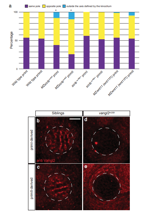
Fig. s3 Vangl2 enrichment analysis in Wild type and the different Wnt and PCP pathway mutants. a. ln all the hair cells that show asymmetric Vangl2 in Figure 3i (green bar), we measured the pole of enrichment with respect to the position of the kinocilium. The results are categorized into three possibilities: asymmetric in the same pole as the kinocilium, asymmetric in the opposite pole than the kinocilium, and asymmetric but outside of the axis defined by the kinocilium. We observe differences between MZscrib and their siblings (Fisher?s exact test p-val WT priml vs MZscrib priml= 0.09724; p-val scrib siblings priml vs MZscrib priml= 0.0828S; p-val WT primll vs MZscrib primll= 0.02S68; p-val scrib siblings primll vs MZscrib primll= 0.03SS7). Wild type and MZwnt11 (wnt11f1) mutants do not show differences (p-val WT priml vs MZwnt11 (wnt11f1) priml= 1; p-val WT primll vs MZwnt11 (wnt11f1) primll= 0.439). b-e. Vangl2 antibody staining on vang/2 siblings priml (b) and primll (c). Vangl2 antibody staining is absent in priml (d) and primll (e) of vang/2 mutants. * denotes statistical significance. Scale bar equals 5Ám.
Image
Figure Caption
Acknowledgments
This image is the copyrighted work of the attributed author or publisher, and
ZFIN has permission only to display this image to its users.
Additional permissions should be obtained from the applicable author or publisher of the image.
Full text @ Nat. Commun.

