IMAGE

Fig. 3

ID
ZDB-IMAGE-200611-3
Source
Figures for Cumplido et al., 2020
Image
Figure Caption

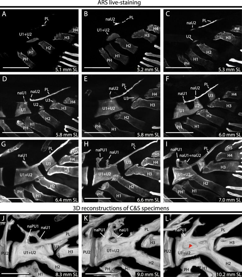
Fig. 3

Mineralization of the compound terminal vertebra. A–I. Zebrafish larvae incubated in Alizarin red S and visualized through confocal microscopy. J–L. 3D reconstructions of cleared and stained individuals visualized through confocal microscopy. Red arrowhead points to the lateral middle ridge or crest. Terminology: cU1 + 2, ural chordacentrum 1 + 2; cU1–3, ural chordacentra 1 to 3; H1–5, hypurals 1 to 5; haPU1, haemal arch of the preural centrum 1; naU1-U2, neural arch of the ural centra 1 or 2; PH, parhypural; PL, pleurostyle. Scale bar: 100 μm

Acknowledgments
This image is the copyrighted work of the attributed author or publisher, and ZFIN has permission only to display this image to its users. Additional permissions should be obtained from the applicable author or publisher of the image. Full text @ Front. Zool.