IMAGE

Fig. 6.

ID
ZDB-IMAGE-200608-21
Source
Figures for Ates et al., 2020
Image
Figure Caption

Fig. 6.

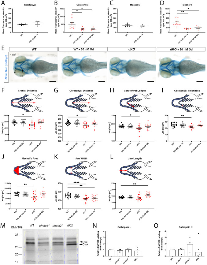
Craniofacial deficits are rescued by Od-mediated inhibition of cathepsin K. (A-D) Mean fluorescence intensity of Col2 immunostaining in the ceratohyal (WT in A, dKO in B) and Meckel's cartilage (WT in C, dKO in D) of 4 dpf larvae with and without Od treatment. (E) Representative images of larvae stained with Alcian Blue. Scale bars: 200 µm. (F-L) Craniofacial morphological measurements at 4 dpf. The measured parameters are highlighted in red in the schematics. (M) In-gel analyses of BMV109, showing cathepsin activities in WT and pheta1/2 mutants at 4 dpf. Blue lines indicate the lane boundaries. (N,O) Quantitation of the cathepsin K and cathepsin L bands from four experiments. Error=s.e.m. ch, ceratohyal; Ctsk, cathepsin K; Ctsl, cathepsin L; m, Meckel's cartilage. *P<0.05, **P<0.01, ****P<0.0001.

Figure Data
Acknowledgments
This image is the copyrighted work of the attributed author or publisher, and ZFIN has permission only to display this image to its users. Additional permissions should be obtained from the applicable author or publisher of the image. Full text @ Dis. Model. Mech.