Image
Figure Caption

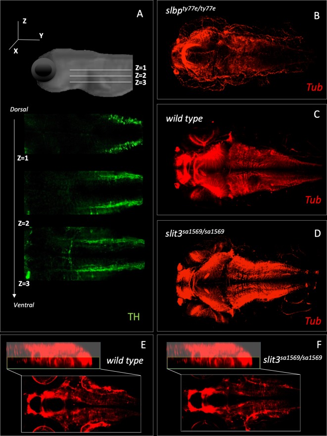
Figure 5-figure supplement 1. Fluorescent immunohistochemistry in three-day old wild type and homozygous mutant <italic>slit3<sup>sa1569</sup></italic> labelled with tyrosine hydroxylase (<bold>A</bold>) and tubulin (<bold>B–F</bold>).

(A) Example of three Z-planes used to quantify catecholaminergic projections crossing the midline. (Stacks 1–3). (B) Anti-tubulin staining of slbpty77e/ty77e mutant larvae, known to have fewer neurons and axonal defects (Turner et al., 2019). (C–D) Anti-tubulin staining of wild type and homozygous mutant slit3sa1569. (E–F) Zoom-in of C-D for midline in the ventral forebrain, where slit3 is known to be expressed (Miyasaka et al., 2005).

Acknowledgments
This image is the copyrighted work of the attributed author or publisher, and ZFIN has permission only to display this image to its users. Additional permissions should be obtained from the applicable author or publisher of the image. Full text @ Elife