IMAGE

Fig. 2

ID
ZDB-IMAGE-200325-51
Antibodies
Source
Figures for Ren et al., 2020
Image
Figure Caption

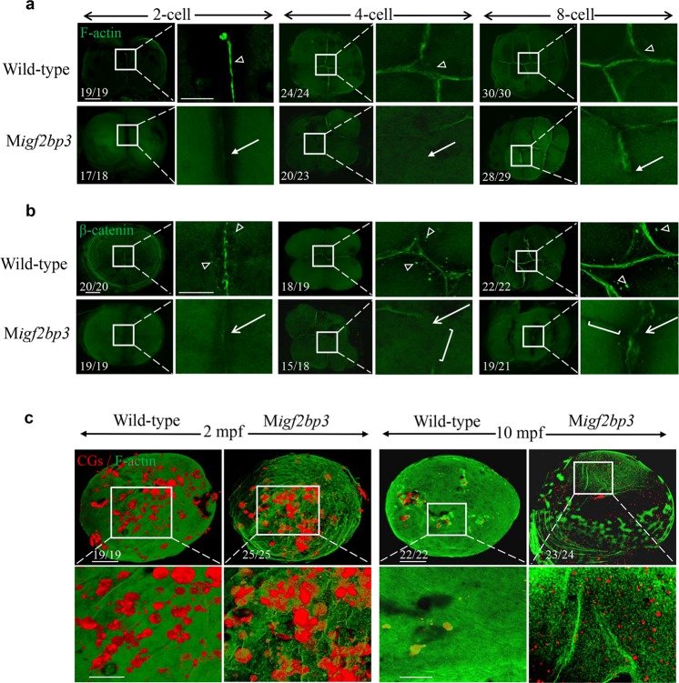
Fig. 2 M<italic>igf2bp3</italic> embryos exhibit abnormal cytoskeleton organization.

a, b Expression and distribution of F-actin and β-catenin in wild type and Migf2bp3 embryos at 2-, 4-, and 8-cell stages. The F-actin and β-catenin were enriched in mature and apparent cleavage furrow (triangle). The inset showed a zoom-in of the boxed region at each stage. Zoomed-out scale bar = 100 μm; zoomed-in scale bar = 50 μm. a Migf2bp3 embryos at 4-cell stage did neither exhibit intact adhesion junction of F-actin cables nor form the mature furrow (white arrow). b Migf2bp3 embryos exhibited reduced expression of β-catenin (white arrows) and did not form intact cell adhesion junction with defective furrow (white bracket). c Double localization of F-actin (green fluorescence) and CG (red fluorescence). The inset showed a zoom-in of the boxed region at each stage. Zoomed-out scale bar = 100 μm; zoomed-in scale bar = 50 μm.

Figure Data
Acknowledgments
This image is the copyrighted work of the attributed author or publisher, and ZFIN has permission only to display this image to its users. Additional permissions should be obtained from the applicable author or publisher of the image. Full text @ Commun Biol