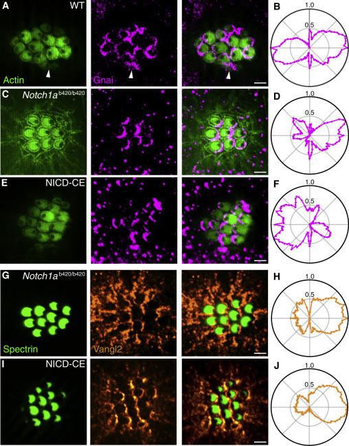
Fig. 4 Parallel Action of Notch and PCP (A) In an apical view of a wild-type neuromast, hair-cell polarity is delineated by actin-GFP (green, left) and Gnai is immunolabeled (magenta, center). As shown in the merged view (right), Gnai already displays a polarized location in immature hair cells (arrowheads). (B) In the average radial intensity profile of Gnai from 239 hair cells of both polarities, caudad and rostrad cells create symmetrical lobes on the two halves of the apical surface. (C) Notch1ab420/b420 hair cells display a consistently caudad orientation with crescents of Gnai at their posterior boundaries. (D) A polar plot from 81 hair cells confirms the asymmetrical distribution of Gnai. (E) NICD-CE hair cells are consistently polarized in the rostrad direction and bear Gnai at their anterior edges. (F) A polar plot from 35 hair cells confirms the asymmetry of Gnai distribution. (G) Immunolabeling for spectrin (green, left) shows that Notch1ab420/b420 hair cells are caudad polarized; labeling for Vangl2 (orange, center) displays its distribution at the posterior edges of the cells. (H) A polar plot from 155 hair cells confirms the asymmetrical distribution of Vangl2. (I) Although NICD-CE hair cells are consistently rostrad-polarized, they also bear Vangl2 at their posterior edges. (J) A polar plot from 235 hair cells confirms the asymmetry. Scale bars, 2 ?m. See also Figure S4.
Image
Figure Caption
Figure Data
Acknowledgments
This image is the copyrighted work of the attributed author or publisher, and
ZFIN has permission only to display this image to its users.
Additional permissions should be obtained from the applicable author or publisher of the image.
Full text @ Curr. Biol.

