IMAGE

Fig. 7

ID
ZDB-IMAGE-200306-57
Antibodies
Source
Figures for Lee et al., 2020
Image
Figure Caption

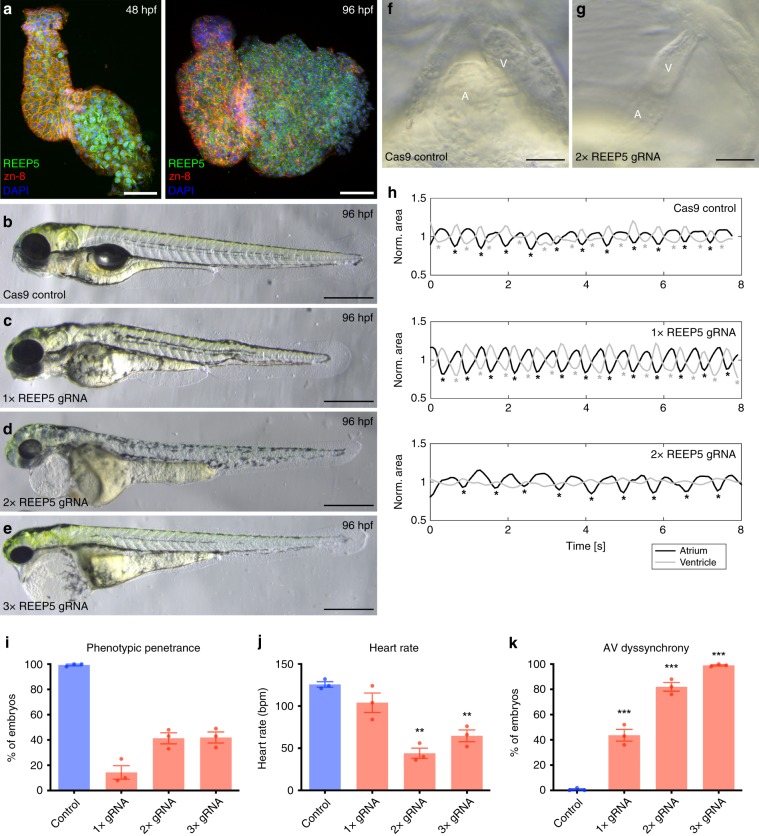
Fig. 7 In vivo CRISPR/Cas9-mediated REEP5 depletion in zebrafish embryos leads to cardiac abnormalities.

a Immunofluorescence analysis of REEP5 expression in wild-type zebrafish hearts at 48 and 96 hpf. Co-staining for REEP5 (green), zn-8 (general plasma membrane; red) and DAPI (nuclear; blue) revealed REEP5 expression in both ventricular and atrial cardiac myocytes, with no nuclear or membrane localization evident. Scale, 50 μm. be CRISPR/Cas9-mediated depletion of REEP5 with varying gRNA concentrations reveals the contribution of REEP5 to embryonic heart development in zebrafish embryos. Images shown are representative of 331 control, 410 2× REEP5 gRNA, and 296 3× REEP5 gRNA total injected embryos, n = 3 independent biological replicates. Scale, 500 μm. f, g Optical imaging analysis of control and 2× REEP5 gRNA injected embryos shows cardiac looping defects associated with REEP5-targeted gRNA injection. Scale, 10 μm. h Movie analysis of REEP5 gRNA injected embryos shows dyssynchronous atrio-ventricular beating rhythms compared to control zebrafish embryos. Area profiles were smoothened with a Gaussian curve with σ = 0.2 s. ik Bar graphs showing i phenotypic penetrance represented as percentage, j heart rate represented as beats per minute (bmp), and k atrioventricular beating dyssynchrony represented as percentage from 331 control, 410 2× REEP5 gRNA, and 296 3× REEP5 gRNA total injected embryos, n = 3 independent biological replicates. Asterisks indicate a statistically significant p value in a Tukey’s multiple comparison analysis where *p < 0.05, **p < 0.01, and ***p < 0.001; data are presented as mean ± SEM.

Figure Data
Acknowledgments
This image is the copyrighted work of the attributed author or publisher, and ZFIN has permission only to display this image to its users. Additional permissions should be obtained from the applicable author or publisher of the image. Full text @ Nat. Commun.