
|
Fig. 3
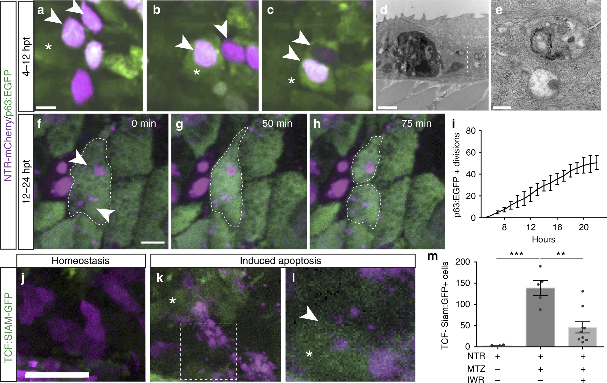
Apoptotic body uptake stimulates Wnt signaling and stem cell division. a?c Confocal maximum intensity projections from time-lapse imaging of a p63-positive cell (green), asterisk, engulfing two apoptotic bodies (magenta), arrowheads (scale?=?5?�m), see Supplementary Movie 2. d, e Transmission electron micrographs of apoptotic bodies in adjacent basal cells (scale?=?2?�m, 500?nm). f?h Time-lapse imaging of a dividing p63:EGFP-positive stem cell that has engulfed apoptotic bodies (arrowheads; scale?=?5?�m), see Supplementary Movie 3. i Quantitation of actively dividing p63:EGFP-positive epithelial stem cells over time after induced apoptosis. j?l Confocal images of Wnt-responsive cells (TCF-Siam:GFP positive) after apoptosis. Arrowheads denote increased Wnt activity in a cell engulfing apoptotic bodies (scale?=?50?�m). m Mean number of TCF-Siam:GFP-positive cells from individual larvae after stem cell ablation ( n?=?5) and treatment with the Wnt inhibitor IWR-1 ( n?=?9). Data are from three independent experiments and error bars represent sem; *** p?<?0.0001, ** p?<?0.0007. One-way analysis of variance (ANOVA) with Dunnett?s mutiple comparisons test (m)