IMAGE

Figure 9.

ID
ZDB-IMAGE-200210-47
Source
Figures for Nagashima et al., 2019
Image
Figure Caption

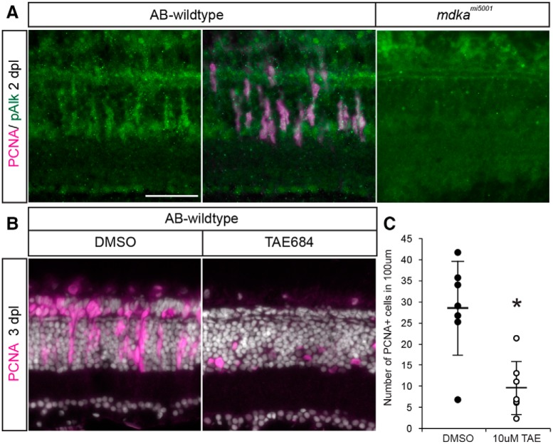
Figure 9.

Activation of the ALK receptor is required for Müller glial to proliferate. A, Double immunostaining for PCNA (magenta) and pAlk (green). PCNA+ cells express pAlk in WT retina at 2 dpl, whereas pAlk immunostaining was not detected in mdkami5001 retina. Scale bar, 30 μm. B, Pharmacological inhibition of Alk using TAE684 suppresses proliferation in the WT retinas after a lesion. C, Counts of PCNA+ proliferative cells in DMSO- or TAE684-treated WT retinas at 3 dpl. n = 7. *p = 0.0011 (nonparametric Mann-Whitney-Wilcoxon).

Figure Data
Acknowledgments
This image is the copyrighted work of the attributed author or publisher, and ZFIN has permission only to display this image to its users. Additional permissions should be obtained from the applicable author or publisher of the image. Full text @ J. Neurosci.