IMAGE

Figure 3.

ID
ZDB-IMAGE-200210-42
Source
Figures for Nagashima et al., 2019
Image
Figure Caption

Figure 3.

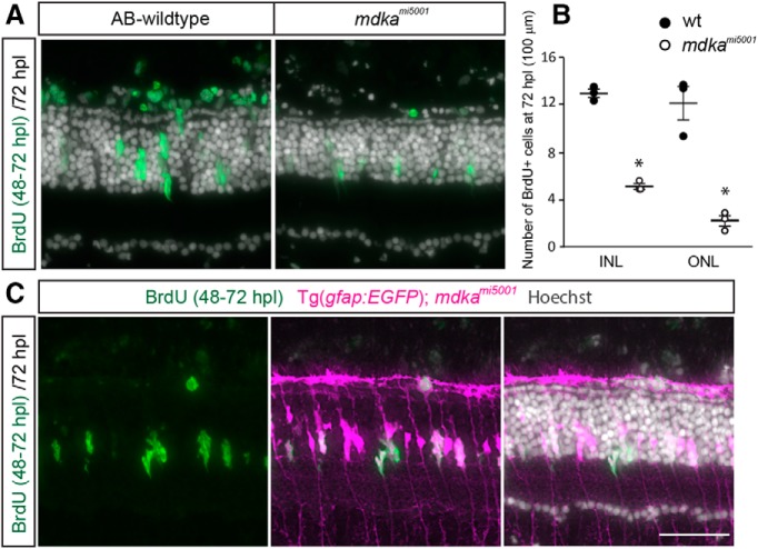
Some Müller glia in the mdkami5001 progress through the cell cycle. A, B, BrdU labeling between 48 and 72 hpl in WT and mdkami5001 mutant retinas. In WT, Müller glia-derived progenitors are labeled with BrdU. In mdkami5001 mutant at 72 hpl, fewer cells in the inner nuclear layer (INL) and outer nuclear layer (ONL) are labeled with BrdU compared with WT (INL: p < 0.0001; ONL: p = 0.0008, ANOVA with post hoc Tukey). C, BrdU labeling (green) in transgenic reporter line, Tg(gfap:eGFP)mi2002, in the mdkami5001 mutant background. Tg(gfap:eGFP)mi2002 is pseudocolored in magenta. Scale bar, 30 μm. *p < 0.01.

Figure Data
Acknowledgments
This image is the copyrighted work of the attributed author or publisher, and ZFIN has permission only to display this image to its users. Additional permissions should be obtained from the applicable author or publisher of the image. Full text @ J. Neurosci.