IMAGE

Fig. 7

ID
ZDB-IMAGE-200207-7
Source
Figures for Tian et al., 2020
Image
Figure Caption

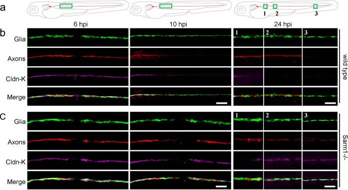
Fig. 7 Schwann cells maintain terminal phenotype in Sarm1-deficient zebrafish.

ac Schematic model of the confocal imaging locations on severed axons (a). Confocal images of wild type (b) and Sarm1−/− (c) specimens in 6, 10, and 24hpi. hpi hour post-injury. The specimens were the Tg[gSAGFF202A;UAS:EGFP; SILL:mCherry] lines and were stained with Claudin-k (magenta) antibody. Scale bar 50 μm.

Acknowledgments
This image is the copyrighted work of the attributed author or publisher, and ZFIN has permission only to display this image to its users. Additional permissions should be obtained from the applicable author or publisher of the image. Full text @ Commun Biol