IMAGE

Figure 4

ID
ZDB-IMAGE-191230-993
Source
Figures for Suzuki et al., 2019
Image
Figure Caption

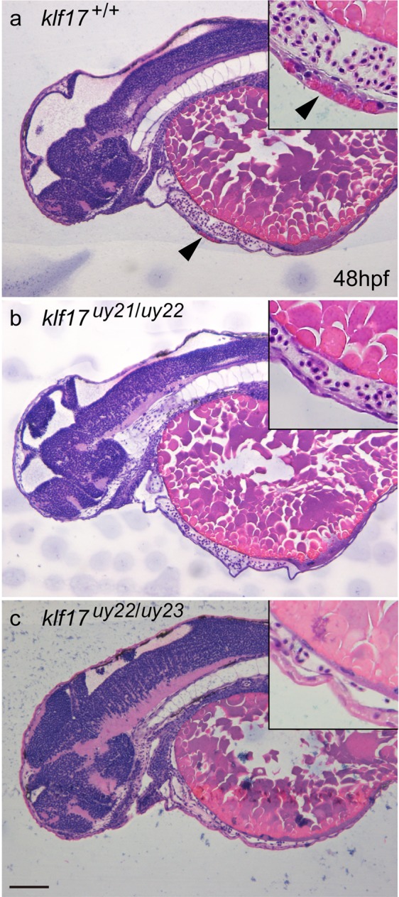
Figure 4

Loss of hatching gland cells in the klf17-deficient embryos. (ac) Cross sections of haematoxylin and eosin (HE)-stained embryos at 48 hpf: wild-type (klf17+/+) (a) klf17-deficient embryos: klf17uy21/uy22 (b) and klf17uy22/uy23. (c) Hatching gland cells observed in wild-type embryo were missing in the klf17-deficient embryos. Arrowheads indicate the position of hatching gland cells. Genomic DNA was isolated from individual caudal fins, with genotyping was performed by genomic PCR. Scale bar, 100 μm.

Figure Data
Acknowledgments
This image is the copyrighted work of the attributed author or publisher, and ZFIN has permission only to display this image to its users. Additional permissions should be obtained from the applicable author or publisher of the image. Full text @ Sci. Rep.