IMAGE

Fig. 5.

ID
ZDB-IMAGE-191230-1890
Source
Figures for Liu et al., 2019
Image
Figure Caption

Fig. 5.

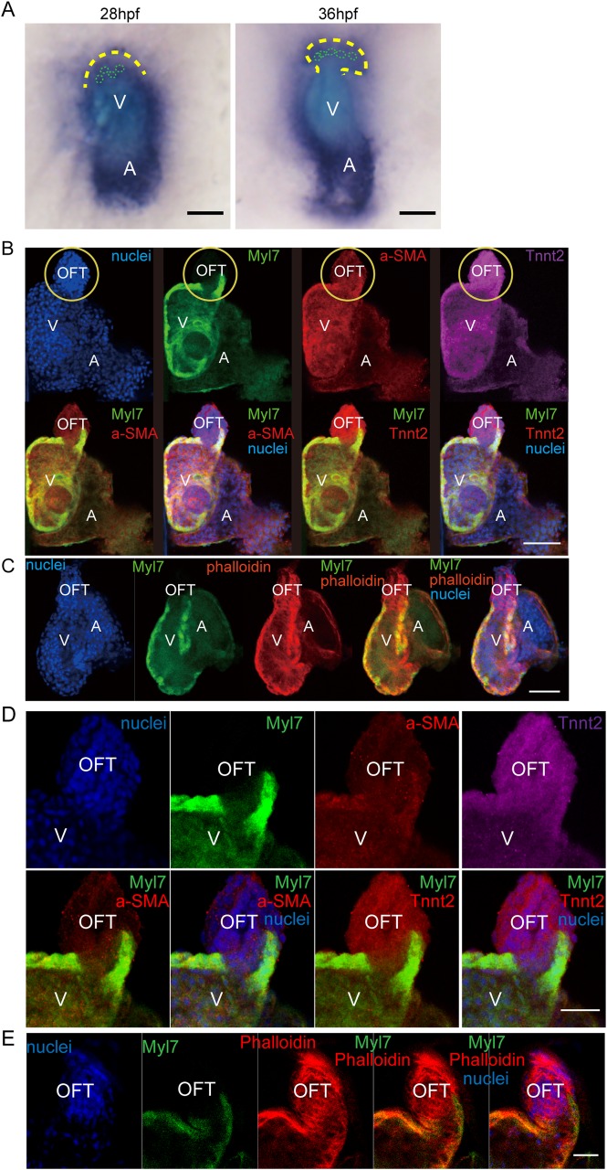
Identification of the TNNT2-positive, Myl7-negative smooth muscle cells in the OFT. (A) Images of in situ hybridization with tnnt2a anti-sense probe. Yellow dotted line, outflow tract outline; green dotted line, cell outline. (B) Immunofluorescence of Tnnt2, Myl7, and α-SMA staining in wild-type zebrafish hearts at 5 dpf. Nuclei is stained by Hoechst 33342. Yellow circle shows OFT area. (C) Myl7 zebrafish heart stained by phalloidin (F-actin) at 5 dpf. Nuclei is stained by Hoechst 33342. Scale bar: 150 µm. (D,E) Images of a single plane of Tnnt2, Myl7, α-SMA and phalloidin immunofluorescence staining. V, ventricle; A, atrium. Scale bars: (A–C) 150 µm, (D,E) 50 µm.

Acknowledgments
This image is the copyrighted work of the attributed author or publisher, and ZFIN has permission only to display this image to its users. Additional permissions should be obtained from the applicable author or publisher of the image. Full text @ Biol. Open