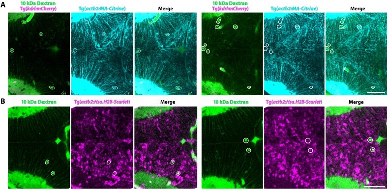
Injected tracers are taken up by parenchymal cells.
(A) Maximum intensity projections (3 µm thick) of the midbrain of membrane labeled (turquoise; Tg(actb2:MA-Citrine) transgenic 3 dpf fish after 10 kDa Dextran injection (green) reveals several parenchymal cells outside of the vasculature (magenta) that contain Dextran within the cell membrane (outlined in white). (B) Maximum intensity projections (5 µm thick) of the midbrain of nuclear labeled (magenta; Tg(actb2:Has.H2B-Scarlet) transgenic fish after 10 kDa Dextran injection (green) reveals several parenchymal cells that are double labeled with Dextran and nuclear signal (outlined in white), further indicating that injected tracers are taken up by select parenchymal cells. Scale bars represent 50 µm.
Acknowledgments
This image is the copyrighted work of the attributed author or publisher, and
ZFIN has permission only to display this image to its users.
Additional permissions should be obtained from the applicable author or publisher of the image.
Full text @ Elife
Your Input Welcome
Thank you for submitting comments. Your input has been emailed to ZFIN curators who may contact you if
additional information is required.
Oops. Something went wrong. Please try again later.