IMAGE

Figure 2 supplement 2.

ID
ZDB-IMAGE-191230-1799
Source
Figures for Gancz et al., 2019
Image
Figure Caption

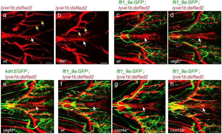
Figure 2 supplement 2. VFL develops normally in lymphatic-related mutants.

(a–h) ventral view of 22dpf (fish size 5–7 mm) fish. (a,b) while medial facial lymphatics are shorter in Tg(lyve1b:dsRed2);flt4-/- as compared to wt siblings (yellow arrows), VFL forms normally (white arrow). Normal VFL is also detected in vegfc+/- heterozygous (c,d), vegfd-/- (e,f) cxcr4a-/- (g) and cxcl12b-/- mutants (h) as compared to wt siblings or age-matched control. Scale bars are 50 µm.

Figure Data
Acknowledgments
This image is the copyrighted work of the attributed author or publisher, and ZFIN has permission only to display this image to its users. Additional permissions should be obtained from the applicable author or publisher of the image. Full text @ Elife