IMAGE

Fig. 6

ID
ZDB-IMAGE-191230-1537
Source
Figures for Xiao et al., 2019
Image
Figure Caption

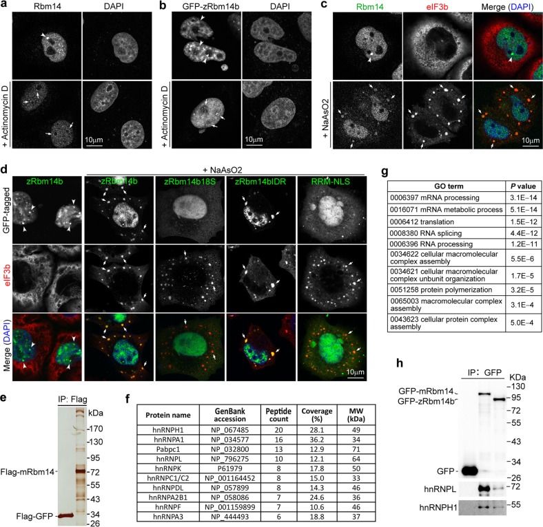
Fig. 6 Rbm14 partitions into RNP compartments and complex with RNA-binding proteins important for RNA metabolism.

a, b The nucleoplasmic puncta (arrowheads) of endogenous human Rbm14 and exogenous zRbm14b were sensitive to RNA pol II activity. HeLa cells that were untransfected (a) or transiently transfected to express GFP-zRbm14b (b) were treated with actinomycin D for 60 min prior to fixation. Nuclear DNA was visualized with DAPI. Arrows indicate fluorescent signals at nucleolar caps. c Human Rbm14 localized to stress granules (SGs; arrows). HeLa cells were treated with sodium arsenite to induce SGs. Untreated cells were used as control. eIF3b served as an SG marker. d zRbm14b translocated into SGs (arrows) through phase separation. HeLa cells transiently expressing the indicated zRbm14b mutants were treated with sodium arsenite. e Mouse Rbm14 associated with large protein complexes. Flag-GFP and Flag-mRbm14 expressed in mouse embryonic stem cells were immunoprecipitated using anti-Flag resin. The immunoprecipitates were resolved by SDS-PAGE and silver stained. f The top 10 hits of potential Rbm14-associated proteins. The immunoprecipitates of Flag-GFP and Flag-Rbm14 were subjected to shotgun mass spectrometric analysis. The top ten hits identified exclusively from the latter sample, according to total peptide count, are listed. g Top ten gene ontology (GO) term hits on candidate Rbm14-associated proteins. Only proteins identified exclusively in the Flag-mRbm14 sample were used for the analysis. h zRbm14b also associated with hnRNP proteins. GFP-tagged mRbm14 and zRbm14b expressed in HeLa cells were immunoprecipitated using anti-GFP resin and immunoblotted to detect the indicated proteins

Acknowledgments
This image is the copyrighted work of the attributed author or publisher, and ZFIN has permission only to display this image to its users. Additional permissions should be obtained from the applicable author or publisher of the image. Full text @ Cell Discov