
Figure 6—figure supplement 1.
- ID
- ZDB-IMAGE-191230-115
- Source
- Figures for Harrison et al., 2019
|
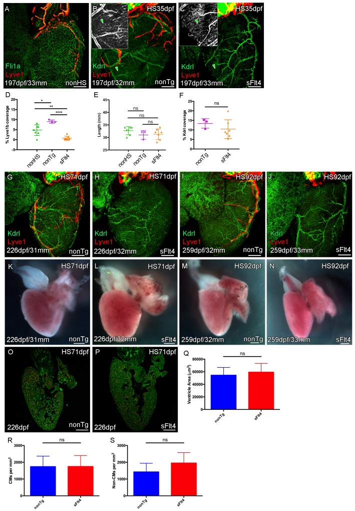
Figure 6—figure supplement 1. Juvenile Vegfc signaling is required for cardiac lymphatic development. Whole-mount confocal and reflected brightfield imaging of adult transgenic zebrafish hearts expressing the pan-endothelial fli1a:GFP (green: A), arterially and endocardially enriched endothelial marker kdrl:GFP (green: B, C, G-J; green arrowhead: B and C), lymphatic endothelial marker lyve1:RFP (red: A-C, G-J). (A) Non-heat shock control (nonHS) zebrafish raised in otherwise identical conditions show normal cardiac lymphatic growth on the ventricle. (B) Increased ventricle lymphatic vasculature in response to heat shock (from 1 m/35dpf) in non-hsp70l:sflt4 transgenic zebrafish (nonTg). (C) Loss of cardiac lymphatic outgrowth onto the ventricle in heat-shocked hsp70l:sflt4 transgenic zebrafish (sFlt4) with no discernable effect on the coronary vessels (green arrowheads) (D) Quantification of lymphatic vessel coverage on the ventricle shows significant changes in coverage; increased in non- hsp70l:sflt4 (n = 3) and decreased in hsp70l:sflt4 (n = 7) fish heat shocked from 35dpf compared to non-heat shocked controls (n = 7; *p=0.0311; **p=0.0028; ****p<0.0001). (E) Heat shocked fish are not significantly (ns) different in length to non-heat shocked controls or each other. (F) Quantification showing there is no significant difference in ventricle coverage of kdrl:GFP with sFlt4 induction. (G and I) Non-transgenic zebrafish have extensions of the cardiac lymphatic vessel (red) onto the ventricle with heart shock from 71 (G) and 92dpf (I). (H and J) Hearts from hsp70l:sflt4 zebrafish heat shocked from 71 (H) and 92dpf (J) completely lack significant extension of cardiac lymphatic vessels onto the ventricle, but show no defects in the coronary vasculature. (K-N) Reflected brightfield images of the same hearts shown in G-J suggest no effect on gross morphology or heart size with heat-shock nonTg (K and M) or heat-shock-induced sFlt4 (L and N). (O and P) Examples images of developmentally heat-shocked (HS 71dpf) hsp70l:sflt4 and nonTg zebrafish with labeled cardiomyocytes (CM, red) and DAPI (green). (Q-S) Quantification of ventricle tissue area (Q), cardiomyocytes number (R), and non-cardiomyocytes number (S) show no significant difference with induction of the sFlt4 transgene in juvenile (HS 71dpf) zebrafish.