
|
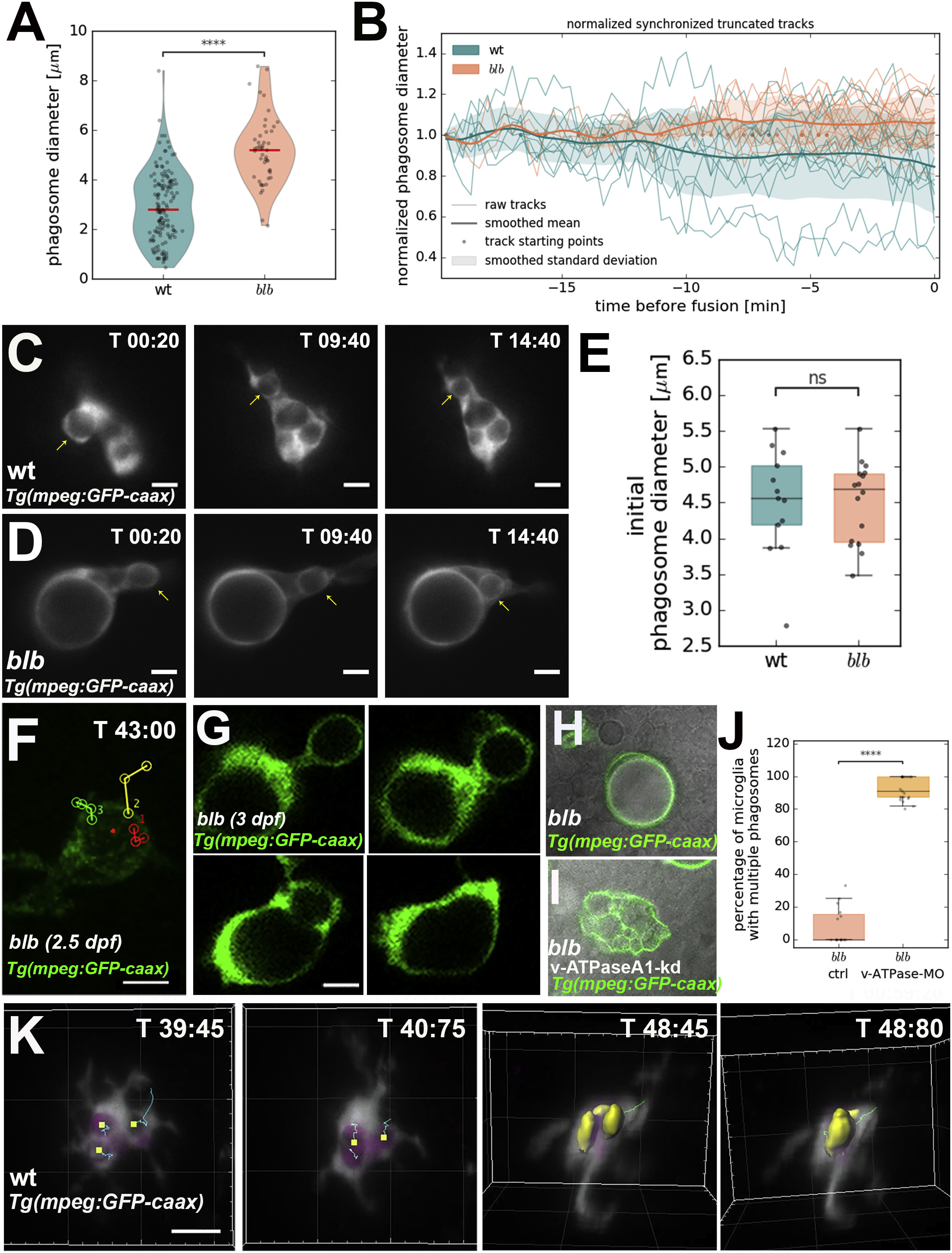
Fig. 4 Tracking of Phagosomes Reveals that These Shrink and Converge into One Cellular Compartment
(A) V-plot showing size distribution of phagosomes in wild-type (n = 27, n = 141) and blbNY007 (n = 27, n = 47). p value < 0.0001.
(B) Quantification of relative vesicle diameter over time in wild-type (n = 13) and blbNY007 (n = 16). t0 corresponds to first fusion even. Data are normalized against the initial size.
(C and D) Representative time-lapse of phagosomal tracking in wild-type (C) (see also Video S3, upper panel) and blbNY007 (D) (see also Video S3, lower panel). Microglia are labeled with Tg(mpeg:GFP-caax). Scale bar, 5 ?m.
(E) Quantification of the size of newly formed phagosomes in wild-type (n = 13) and in blbNY007 (n = 16). Whiskers 5th/95th percentile, p value: not significant.
(F) SPIM time-lapse of 2.5 dpf blbNY007 microglia labeled with Tg(mpeg:GFP-caax). The red dot marks the growing vesicle. Red, yellow, and blue tracks show the trajectories of three incoming phagosomes. Time indicated in minutes. Scale bar, 10 ?m. See also Video S5.
(G) High time resolution SPIM time-lapse of a representative fusion event in a 3 dpf blbNY007 microglia labeled with Tg(mpeg:GFP-caax). Scale bar, 5 ?m.
(H and I) Representative examples of microglia labeled with Tg(mpeg:GFP-caax) in blbNY007 (H) and in blbNY007 injected with a morpholino against the a1-vATPase (I) to prevent vesicular fusion.
(J) Quantification of experiment in (H) and (I). Percentage of microglia with more than 1 vesicle per cell in blbNY007 (n = 15) and blbNY007 injected with a morpholino against the a1-vATPase (n = 21), whiskers: 5th/95th percentile, p value < 0.0001.
(K) SPIM time-lapse of wild-type microglia labeled in Tg(mpeg::GFP-caax). Tracking highlights fusion events. Scale bar, 5 ?m. Time in minutes. See also Video S4.
Reprinted from Developmental Cell, 49(1), Villani, A., Benjaminsen, J., Moritz, C., Henke, K., Hartmann, J., Norlin, N., Richter, K., Schieber, N.L., Franke, T., Schwab, Y., Peri, F., Clearance by Microglia Depends on Packaging of Phagosomes into a Unique Cellular Compartment, 77-88.e7, Copyright (2019) with permission from Elsevier. Full text @ Dev. Cell