Fig. 4
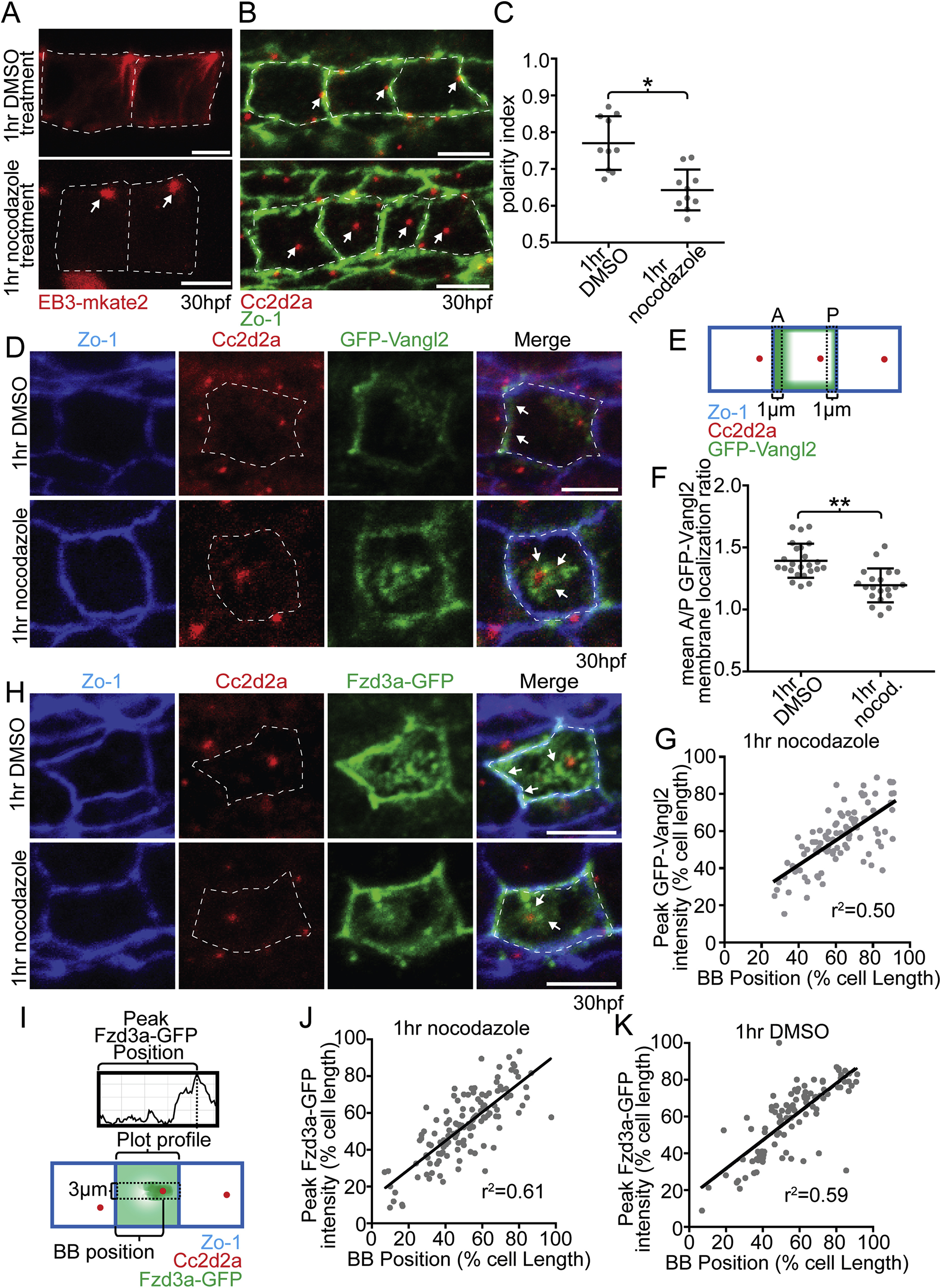
Microtubules are required to maintain floorplate PCP. (A) Live lateral views of floorplate cells expressing Tg(shh:gal4); Tg(uas:EB3-mkate2) at 30 hpf. Upon nocodazole treatment EB3-mkate2 labeled MTs collapse to subapical foci near the BB (arrows). (B) Ventral views of Cc2d2a (red) and Zo-1 (green) immunostaining Arrows indicate position of BBs. Nocodazole treatment disrupts the posterior localization of BBs. (C) Quantitation of per embryo average BB polarity index after 1hr DMSO treatment (control) or 1hr nocodazole treatment. N ?= ?200 ?cells, 20 embryos; *p ?= ?0.0003; significance was determined with Mann-Whitney test. (D) Ventral views of 30hpf Tg(shh:gal4); Tg(uas:GFP-Vangl2) embryos. White dotted lines indicate cell boundaries based on ZO-1 staining. Arrows indicate GFP-Vangl2 localization at the anterior membrane in controls and around the BB in nocodazole-treated embryos. (E) Diagram illustrating how anterior and posterior membrane levels of GFP-Vangl2 were measured in fixed ventral floorplate images. (F) Quantitation of per embryo average GFP-Vangl2 anterior vs. posterior membrane localization ratios in isolated floorplate cells expressing Tg(shh:gal4); Tg(uas:GFP-Vangl2). N ?= ?416 ?cells, 66 embryos; **p ?< ?0.0001, *p ?= ?0.0013; significance was determined with a Kruskal-Wallis test with Dunn's multiple comparison. (G) Graph of the correlation between GFP-Vangl2 and BB localization. After 1hr nocodazole treatment peak GFP-Vangl2 intensity correlates with the position of the BB N ?= ?10 embryos, 100 ?cells, r2?=?0.50. (H) Ventral views of floorplate cells at 30 hpf immunostained for Fzd3a-GFP (green), Zo-1 (blue) and Cc2d2a (red) after 1hr DMSO treatment (control) or 1hr nocodazole treatment. (I) Diagram illustrating how peak Fzd3a-GFP localization and BB positions were measured. BB distance from anterior membrane was measured compared to overall anterior-posterior cell length. A 3?μm wide ROI centered on the position of the BB was drawn from anterior to posterior membranes. The “plot profile” tool in ImageJ was used to measure average GFP levels across the anterior to posterior cell axis. (J,K) Correlation between Fzd3a-GFP localization and BB position in nocodazole-treated and control floorplate cells. Each data point represents the measurements from a single cell. J: N?=?100?cells, 10 embryos; r2?=?0.61; K: N?=?100?cells, 10 embryos; r2?=?0.59. Scale bars: 5?μm.
Reprinted from Developmental Biology, 452(1), Mathewson, A.W., Berman, D., Moens, C.B., Microtubules are required for the maintenance of planar cell polarity in monociliated floorplate cells, 21-33, Copyright (2019) with permission from Elsevier. Full text @ Dev. Biol.

