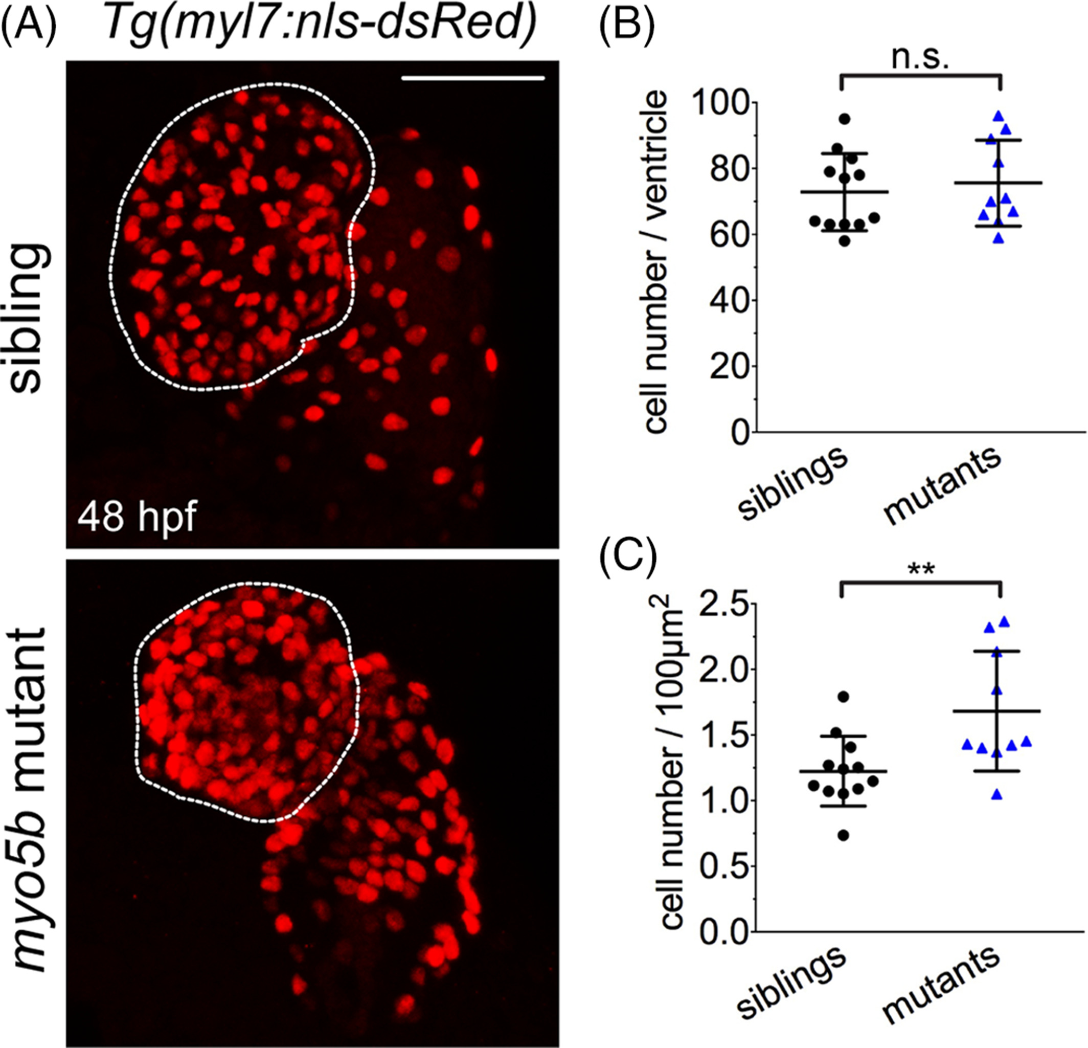
Fig. 5 Cardiomyocyte number is unchanged between siblings and uq 23ks mutants. A: Confocal imaging of sibling and uq 23ks mutants on the Tg(myl7:nls?dsRed) background at 48 hpf. B: Dot plots quantifying the total number of cardiomyocytes observed in embryonic hearts show no significant (ns) difference between sibling and uq 23ks mutants at 48 hpf, suggesting a difference in cell proliferation or differentiation is not contributing to the uq 23ks phenotype. C: Quantification of cell number per area, however, is significantly higher in uq 23ks mutants compared with siblings, consistent with smaller cardiomyocyte size in uq 23ks mutants. mean?±?SEM. n.s. = not significant; **P?<?0.01.
Image
Figure Caption
Figure Data
Acknowledgments
This image is the copyrighted work of the attributed author or publisher, and
ZFIN has permission only to display this image to its users.
Additional permissions should be obtained from the applicable author or publisher of the image.
Full text @ Dev. Dyn.

