
|
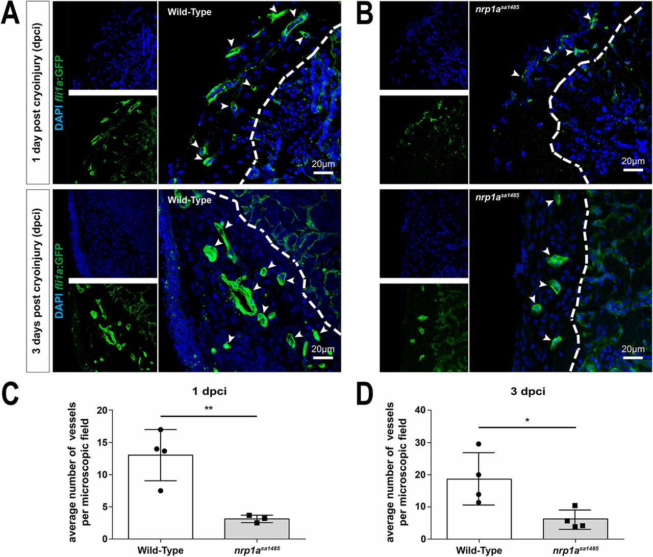
Fig. 5
Neovascularization of the cryoinjured area is impaired in nrp1asa1485 mutants. (A,B) Blood vessels in either wild-type (A) or nrp1asa1485 (B) tg(fli1a:EGFP)y1 zebrafish at 1 (upper row) and 3 (lower row) dpci were identified in heart sections using GFP immunofluorescence in vascular structures. Heart sections were also counterstained with DAPI. Smaller images (left) represent DAPI staining (blue) and GFP staining (vessels, green) only; larger images (right) are the merged images. The white dashed line delineates the border of the area of injury. White arrowheads indicate blood vessels. (C,D) GFP-positive vessels were quantified at 1 dpci (C; **P<0.01, two-tailed t-test of n=3 and 4) and 3 dpci (D; *P<0.05, two-tailed t-test of n=4) for wild-type (white bars) versus nrp1asa1485 (gray bars) hearts. Individual data points (circles for wild type and squares for nrp1asa1485) represent individual hearts, each averaged from vessel counts in three to four different sections covering the injury site.