
|
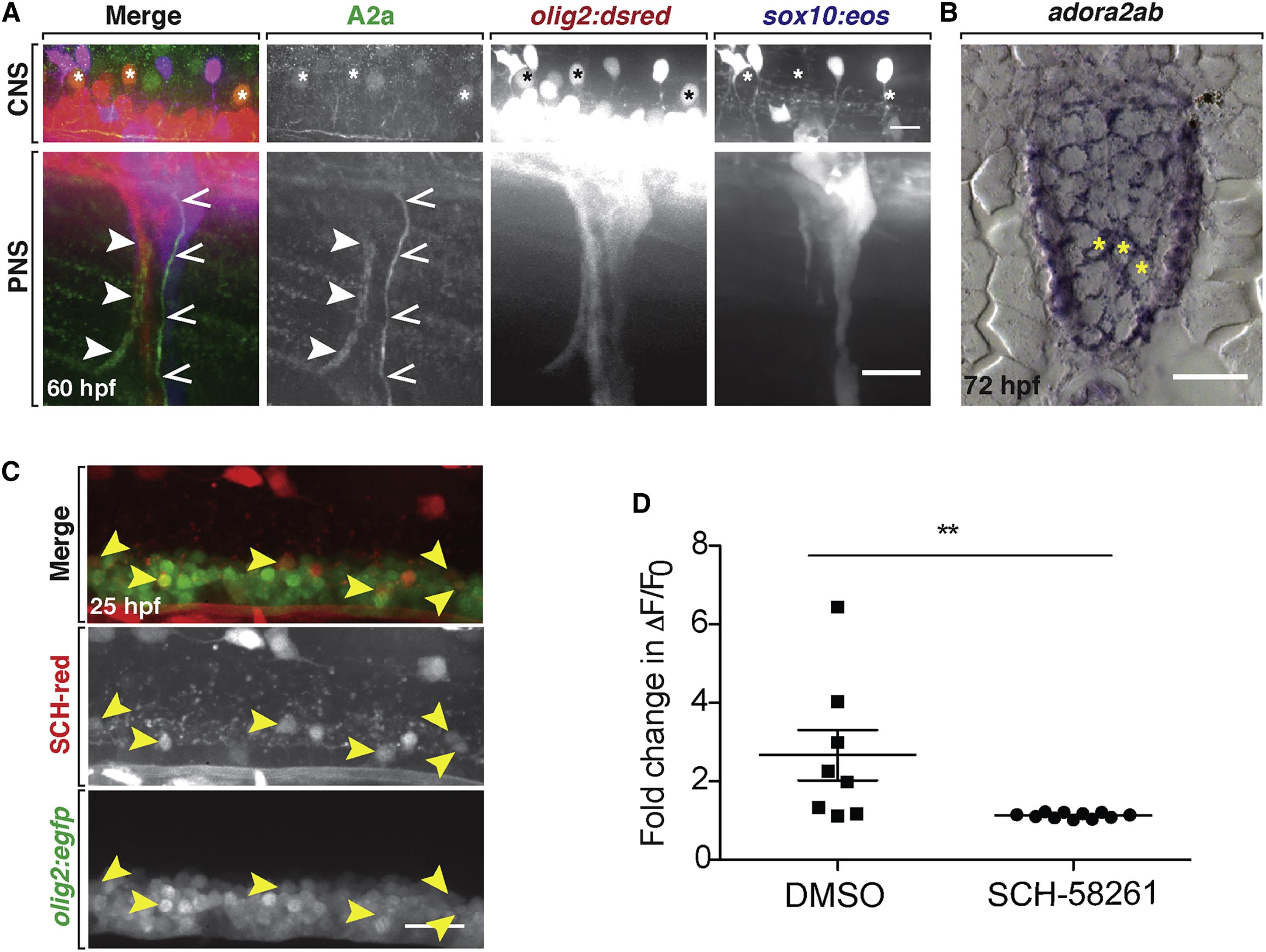
Fig. 5
A2a AR Inhibition Reduces Neural Activity of Spinal Cord Neurons
(A) Lateral views of a 60 hpf WT olig2:dsred;sox10:eos larva stained with an A2a AR antibody. Top: spinal cord (CNS) with A2a expression in a subset of olig2+ motor neurons (asterisks). Bottom (PNS): peripheral spinal nerves with A2a expression in motor (closed arrowheads) and sensory (open arrowheads) axons.
(B) adora2ab mRNA expression at 72 hpf in the spinal cord of a WT larva. Yellow asterisks denote motor neurons.
(C) Twenty-five hours post-fertilization olig2:egfp embryo labeled with SCH-red fluorescent antagonist. Arrowheads mark spinal cord olig2+/SCH-red+ motor neurons.
(D) Mean ± SEM of the fold change in ΔF/F0 over time per individual firing event for larvae treated with DMSO (n = 8 neurons from eight larvae) and SCH-58261 (n = 11 neurons from ten larvae); p = 0.01. Scale bars, 20 μM.