IMAGE

Fig. 3

ID
ZDB-IMAGE-190723-96
Source
Image
Figure Caption

Fig. 3

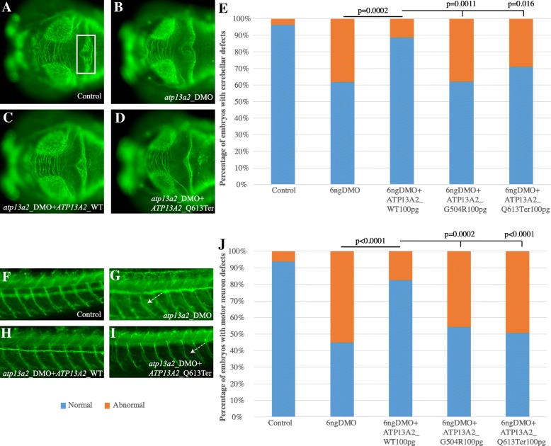
ad Dorsal view of the brain of 3 days post fertilization (dpf) zebrafish larvae, visualized with an anti-acetylated tubulin antibody. The photographs of a control embryo (a), an embryo injected with morpholino (MO) oligonucleotides against exons 4 and 6 of the endogenous atp13a2 (DMO; b), an embryo co-injected for DMO and wild-type (WT) human ATP13A2 (c), and an embryo co-injected for DMO ATP13A2 encoding the p.Glu613Ter change (d). The area of the cerebellum that was evaluated is highlighted with a white box in the control embryo (a). In embryos injected with DMO (b), significant disorganization of the axons that branch in the midline of the cerebellum is noted. The cerebellar phenotype was rescued by co-injection of DMO with WT human ATP13A2 (c), but not with DMO + ATP13A2 p.Glu613Ter (d) or DMO + ATP13A2 p.Gly504Arg (data not shown), suggesting that these represent loss-of-function alleles. d Quantification of the percentage of embryos with cerebellar defects. fi Lateral view of the peripheral nervous system (PNS) of two dpf embryos, showing the motor neurons that extend from the notochord dorsally to innervate the myotomes ventrally. The photographs of a control embryo (f), an embryo injected with DMO (g), DMO + ATP13A2 WT (h), and DMO + ATP13A2 p.Glu613Ter (i). In embryos injected with DMO (g), motor neurons deviate from the normal dorso-ventral path (white arrows). The phenotype was rescued by co-injection of DMO with ATP13A2 WT (h), but not with DMO + ATP13A2 p.Glu613Ter (i). j Quantification of the percentage of embryos with cerebellar defects. For both in vivo complementation assays, statistical significance was determined using a χ2 test

Figure Data
Acknowledgments
This image is the copyrighted work of the attributed author or publisher, and ZFIN has permission only to display this image to its users. Additional permissions should be obtained from the applicable author or publisher of the image. Full text @ Hum. Genomics