IMAGE

Fig. 6.

ID
ZDB-IMAGE-190723-899
Genes
Source
Figures for Cho et al., 2019
Image
Figure Caption

Fig. 6.

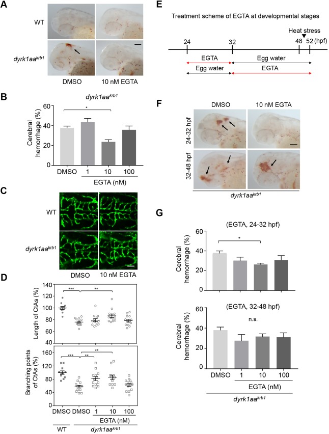
EGTA identified by the chemical library screening effectively rescues the cerebral hemorrhage and abnormal CtA development of dyrk1aakrb1 mutants. (A) o-dianisidine staining images at 52 hpf showing that the cerebral hemorrhage (arrow) of dyrk1aakrb1 embryos were rescued by treating with 10 nM EGTA. WT embryos were not affected by the treatment at the same concentration. (B) Quantitation of the rescue of the cerebral hemorrhagic phenotype by EGTA. The cerebral hemorrhage in dyrk1aakrb1 embryos is reduced from 37.5% to 23.6% by 10 nM EGTA treatment. The data is presented with five independent experiments with approximately 20 embryos in each repeat. (C) Compiled confocal microscopy images of the rescue effect on angiogenic defects of CtAs in dyrk1aakrb1 embryos by EGTA treatment. (D) Quantitative data showing that the reduced mean percentages of length and branching points of CtAs in dyrk1aakrb1 embryos at 52 hpf (76% and 57.4%, respectively, compared to WT embryos as 100%) were increased up to 86.6% and 85.9%, respectively, by 10 nM EGTA treatment. n≥11 each group. (E) Schematic depicts the treatment scheme of EGTA at developmental stages from 24 to 52 hpf in dyrk1aakrb1 embryos. (F) The cerebral hemorrhage of dyrk1aakrb1 embryos at 52 hpf (arrows) were rescued by 10 nM EGTA treatment for 24-32 hpf but not by the treatment for 32-48 hpf. (G) Quantitative data showing that cerebral hemorrhage of dyrk1aakrb1 embryos reduced from 37.7% to 26.3% by treating with 10 nM EGTA for 24-32 hpf. *P<0.05, **P<0.01, ***P<0.005 (one-way ANOVA). n.s., not significant. Data are mean±s.e.m. Scale bars: 100 µm in A,F; 50 µm in C.

Figure Data
Acknowledgments
This image is the copyrighted work of the attributed author or publisher, and ZFIN has permission only to display this image to its users. Additional permissions should be obtained from the applicable author or publisher of the image. Full text @ Dis. Model. Mech.