IMAGE

Figure 10.

ID
ZDB-IMAGE-190723-1792
Source
Figures for Chen et al., 2019
Image
Figure Caption

Figure 10.

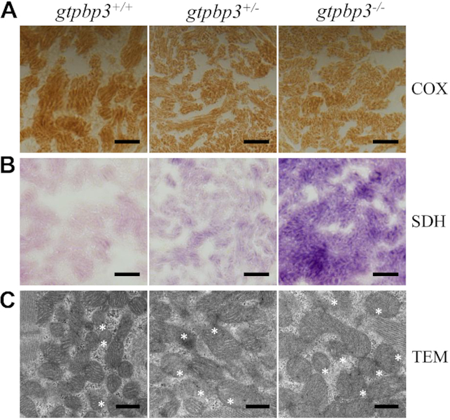
Mitochondrial defects in the zebrafish heart. (A, B) Assessment of mitochondrial function in cardiomyocytes by enzyme histochemistry (EHC) staining for COX (A) and SDH (B) in the frozen-sections of ventricles in the gtbpbp3−/−, gtpbp3+/− and gtpbp3+/+ zebrafish at one year old. Scale bars: 40 μm. (C) Mitochondrial networks from cardiomyocytes of transmission electron microscopy. Ultrathin sections were visualized with 30 000× magnifications.

Acknowledgments
This image is the copyrighted work of the attributed author or publisher, and ZFIN has permission only to display this image to its users. Additional permissions should be obtained from the applicable author or publisher of the image. Full text @ Nucleic Acids Res.