
Image
|
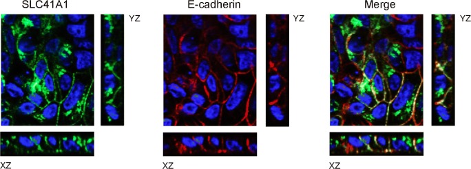
Figure Caption
Fig. 9
SLC41A1 is expressed at the basolateral membrane. Localization of SLC41A1 (green) and E-cadherin (red) in stably transfected polarized MDCKI cells with HA-tagged SLC41A1 is shown by immunofluorescence and confocal analyses in Z, XZ, and YZ sections. In blue, DAPI staining
Acknowledgments
This image is the copyrighted work of the attributed author or publisher, and
ZFIN has permission only to display this image to its users.
Additional permissions should be obtained from the applicable author or publisher of the image.
Full text @ Pfl�gers Archiv. / Eur. J. Physiol.