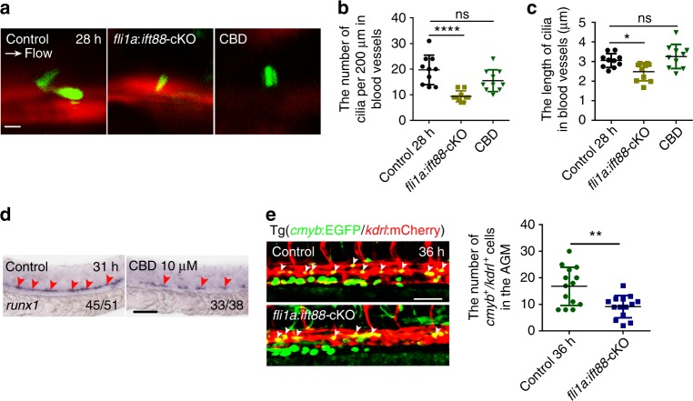
Blocking formation or function of primary cilia impairs hematopoietic stem and progenitor cell (HSPC) development. a Confocal imaging of cilia in the aorta-gonad-mesonephros (AGM) region in kdrl:mCherry/βact:Arl13b–GFP double-transgenic line with fli1a:ift88-cKO-functional injection, or ciliobrevin D (CBD) treatment at 28 hpf. White arrow denotes the blood flow direction. Scale bar, 5 µm. b, c The quantification of primary cilia number and length in the blood vessels of AGM in a. Data in b, c represent the analysis results of one-way ANOVA–Dunnett test. Error bars, mean ± s.d., n = 10 embryos. ns non-significant, *P < 0.05, ****P < 0.0001. d WISH result of runx1 (red arrowheads) in control and CBD-treated embryos at 31 hpf. The red arrowheads indicate the expression of HSPC marker runx1. Scale bar, 100 µm. e The kdrl:mCherry+/cmyb:EGFP+ HE cells (white arrowheads) in the AGM region in fli1a:ift88-cKO-injected embryos (left panel) with quantification (right panel) at 36 hpf. Cmlc2:EGFP-negative embryos were used as a negative control (control). Scale bar, 50 µm. Error bars, mean ± s.d., n = 14 embryos. **P < 0.01, Student’s t-test
This image is the copyrighted work of the attributed author or publisher, and
ZFIN has permission only to display this image to its users.
Additional permissions should be obtained from the applicable author or publisher of the image.
Full text @ Nat. Commun.
Your Input Welcome
Thank you for submitting comments. Your input has been emailed to ZFIN curators who may contact you if
additional information is required.
Oops. Something went wrong. Please try again later.