IMAGE

Figure 2.

ID
ZDB-IMAGE-190723-1044
Source
Figures for Alassaf et al., 2019
Image
Figure Caption

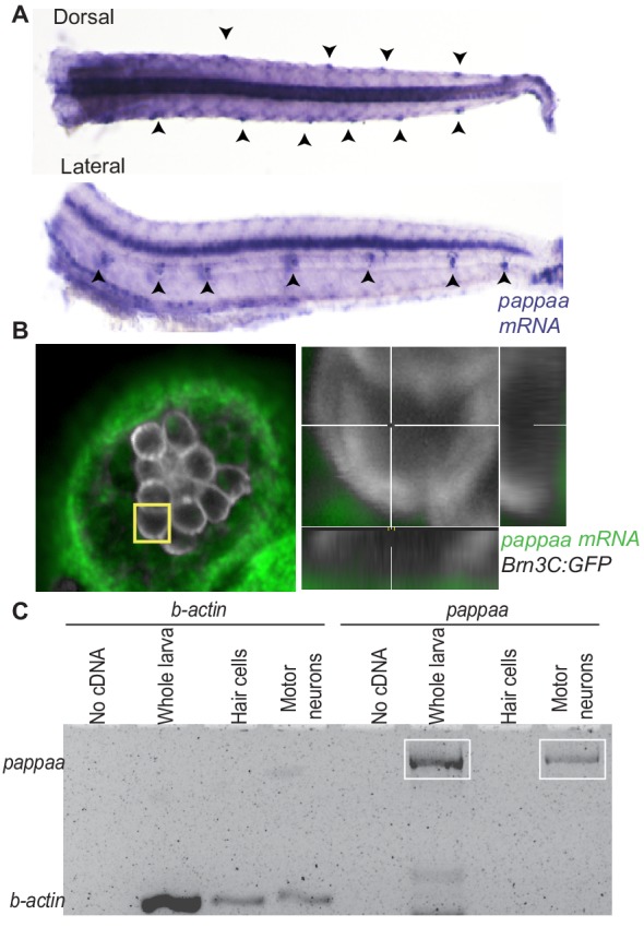
Figure 2. <italic>pappaa</italic> is expressed by neuromast support cells and motor neurons.

(A) Whole mount in situ hybridization shows pappaa mRNA expression at four dpf by lateral line neuromasts (arrowheads). (B) Fluorescent in situ hybridization of pappaa (green) and brn3c:GFP labeled hair cells (white) shows pappaa mRNA expression by the support cells that surround hair cells. (C) RT-PCR of fluorescently sorted brn3c:GFP labeled hair cells and mnx1:GFP labeled motor neurons shows pappaa expression by motor neurons, but not hair cells. RT-PCR products represent pappaa cDNA fragment.

Acknowledgments
This image is the copyrighted work of the attributed author or publisher, and ZFIN has permission only to display this image to its users. Additional permissions should be obtained from the applicable author or publisher of the image. Full text @ Elife