
Image
|
Figure Caption
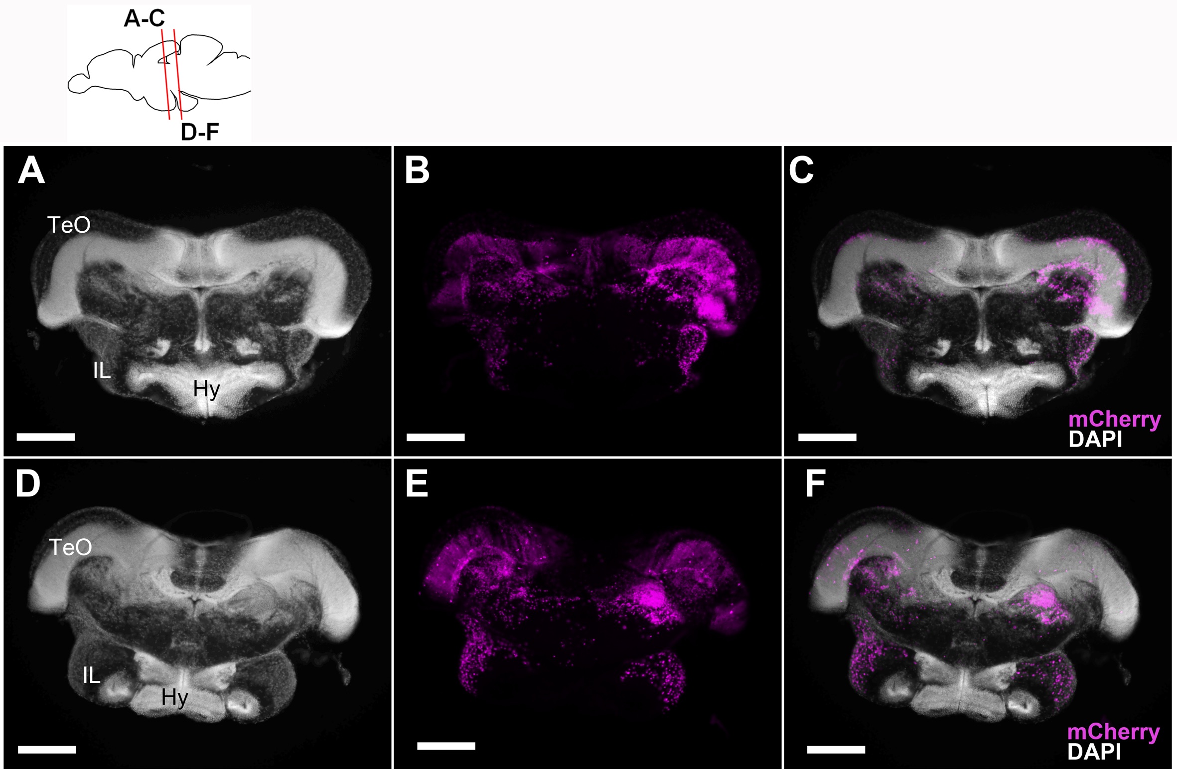
Fig. S2
Localization of the mCherry-positive cells in the 5 wpf juvenile brain of Tg(her5:ERT2CreERT2;βact:lox-stop-lox-hmgb1:mCherry) zebrafish treated with tamoxifen at 24 hpf. Frontal sections showing mCherry-positive cells in magenta and DAPI nuclear labeling in gray. A-C show the anterior IL and D-E show more posterior IL. Scale bars: 100 μm. Abbreviations, Hy: hypothalamus, IL: inferior lobe, TeO: optic tectum. (TIF 10750 kb)
Acknowledgments
This image is the copyrighted work of the attributed author or publisher, and
ZFIN has permission only to display this image to its users.
Additional permissions should be obtained from the applicable author or publisher of the image.
Full text @ BMC Biol.