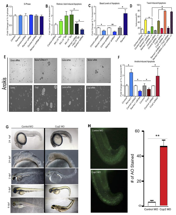
Fig. 2
RARRES1 and CCP2 regulate apoptosis but not cell proliferation. A. Cells were stained with propidium iodide (Sigma) and analyzed by flow cytometry. % S-phase cells are expressed as fold change in MCF10A cells exogenously expressing RARRES1 or following transient or stable knockdown. B. RARRES1 depletion inhibits retinoic acid induced apoptosis in MCF10A cells. C. RARRES1 exogenous expression, transient and stable depletion, regulate basal levels of apoptosis. D. Taxol-induced cell apoptosis. % cell death after RARRES1 or CCP2 manipulation, with, or without taxol (C-control, T-taxol treatment (10ug/ml for 48h). Samples were stained with fluorescein-labeled Annexin V and propidium iodide and analyzed by flow cytometry. E. and F. RARRES1 and CCP2 have reciprocal effect on anoikis-induced apoptosis. Phase-contrast photographs of cell aggregates/spheroids E. and fold change of RARRES1 or CCP2 depleted cells following suspension-induced apoptosis (anoikis) in MCF10A cells F.. G. CCP2 knockdown causes transient cell death in zebrafish embryos. (1-10] Phenotype following injection of 8 ng control MO [1, 3, 5, 7, 9], or 8 ng agbl2 MO [2, 4, 6, 8, 10]. H. Quantification of CCP2 knockdown induced cell death measured by acridine orange staining

