
Image
|
Figure Caption
Fig. 6
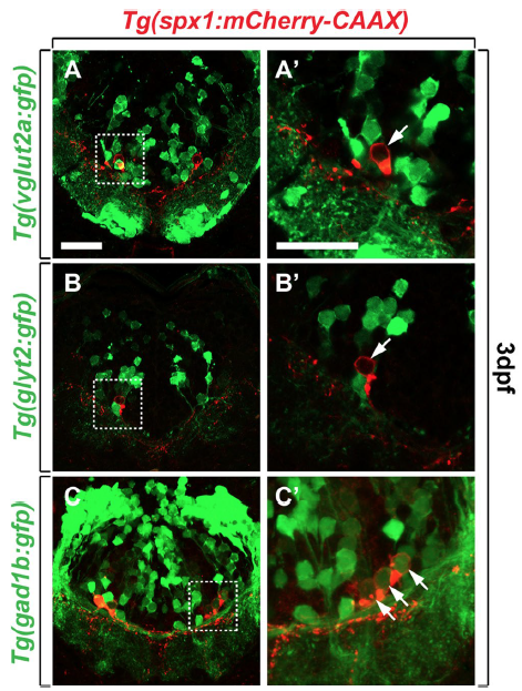
Identification of neurotransmitter phenotype of SPX1 neurons in the hindbrain. (A–C’) Transverse sections of the hindbrain of Tg(spx1:mCherry-CAAX); Tg(vglut2a:gfp) (A,A’), Tg(spx1:mCherry-CAAX); Tg(glyt2:gfp) (B,B’), and Tg(spx1:mCherry-CAAX); Tg(gad1b:gfp) (C,C’) at 3 days post-fertilisation (dpf), dorsal to the top. Panels labelled with a prime are the magnified images of the boxed areas in each panel. Arrows mark vglut2a-negative (A’), glyt2-negative (B’) and gad1b-positive (C’) spx1-expressing neurons in the hindbrain. Scale bar: 25 μm.
Figure Data
Acknowledgments
This image is the copyrighted work of the attributed author or publisher, and
ZFIN has permission only to display this image to its users.
Additional permissions should be obtained from the applicable author or publisher of the image.
Full text @ Sci. Rep.