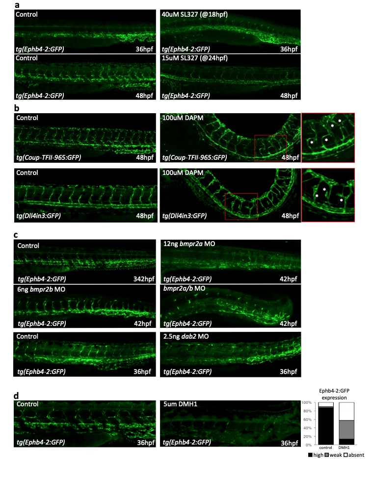
Fig. S12
Perturbation of signalling through Notch, ERK/MAPK and BMPR2 has little effect on the activity of Ephb4-2, while inhibition of ALK1/2/3/6 reduces Ephb4- 2 activity. a. Representative 36 and 48hpf tg(Ephb-2:GFP) zebrafish after DMSO alone (control) or addition of the ERK inhibitor SL327 demonstrate that Ephb4-2:GFP expression does not expand into the artery after ERK inhibition. Although high levels of SL327 inhibit arterial formation, both dorsal aorta and dorsal/ventral veins were clearly visible (using circulation) in fish after 15uM SL327 added at 24hpf. (n=40 for all groups). b. Representative 48hpf tg(Coup-TFII-965) and tg(Dll4in3:GFP) zebrafish after DMSO alone (Coup-TFII n=51, Dll4 n=42 ) or 100uM DAPM treatment (Coup-TFII n=57, Dll4 n=54) demonstrate that Notch inhibition does not change Coup-TFII965:GFP expression intensity or venous localization. As previously reported after Notch pathway inhibition, hypersprouting was clearly visible (red box denotes zoomed region), particularly in the tg(Dll4in3:GFP) zebrafish. c. Representative 42hpf tg(Ephb-2:GFP) zebrafish without treatment (control, picture shown is from bmpr2a injection set) or after injection of bmpr2a MO (control n =30, MO n =32), bmpr2b MO (control =35, MO n= 25), combined bmpr2a/b MO (control n=40, MO n=40) and dab2 MO (control n=51, MO n=45) Ephb4-2:GFP expression was detected in all, although defects in the sprouting from the caudal vein were detected as previously described by 5,6. d. Inhibition of BMP signalling by DMH1 severely reduces the expression of the venous Ephb4-2:GFP transgene in zebrafish. Graph depicts observed expression pattern of GFP in tg(Ephb4-2:GFP) embryos for DMSO-treated control (n=42) and 5µM DMH1-treated embryos (n=68), black represents high expression, grey represents weak, white represents no detected expression. Embryos shown are representative 36hpf tg(Ephb4-2:GFP) embryos after control and DMH1-treatment embryos.

