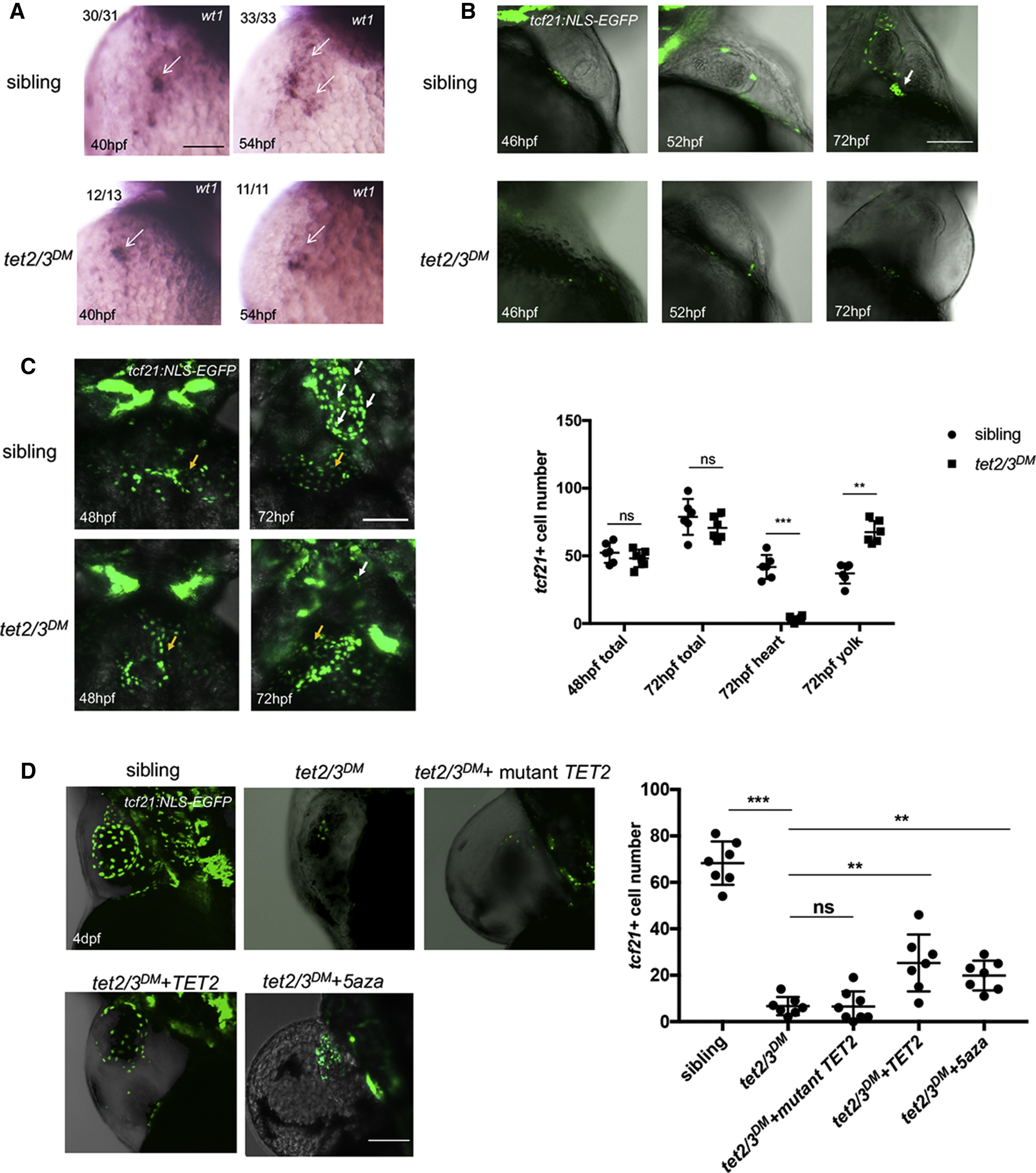
Fig. 1
tet2 and tet3 Have Overlapping Functions in PE Recruitment to the Heart
(A) WISH for PE marker wt1 at 40 hpf and 54 hpf. Arrows indicate PE cells with wt1transcripts.
(B) Lateral view of hearts showing GFP-labeled PE and epicardium in 46-hpf, 52-hpf, and 72-hpf larvae carrying the Tg(tcf21:NLS-EGFP) transgene. White arrow indicates the extracellular matrix bridge between AVC and the pericardial wall.
(C) Ventral view of hearts showing GFP-labeled PE and epicardium in 48-hpf and 72-hpf sibling or tet2/3DM larvae carrying the Tg(tcf21:NLS-EGFP) transgene. White arrows indicate tcf21+ PE and epicardial cells located on the heart. Yellow arrows indicate tcf21+ PE and epicardial cells located on the yolk sac. Graph indicates the total number of PE and epicardial cells in 48-hpf or 72-hpf sibling and tet2/3DMlarvaeand the number of PE and epicardial cells located on heart or yolk sac at 72-hpf sibling and tet2/3DM larvae.
(D) The PE migration defect is partially rescued by TET2 mRNA injection or 5-aza treatment. GFP-labeled PE and epicardium in 4-dpf sibling, tet2/3DM, and tet2/3DMinjected with wild-type hTET2 mRNA, mutant hTET2 mRNA, or tet2/3DM exposed to 75 μM 5-aza larvae carrying the Tg(tcf21:NLS-EGFP) transgene. Graph indicating the number of epicardial cells located on the heart at 4 dpf is shown.
Scale bars: (A) 50 μm; (B–D) 100 μm. ∗∗p < 0.01; ∗∗∗p < 0.001; ns indicates not significant. Data are presented as mean ± SD derived from at least three independent biological replicates

