
|
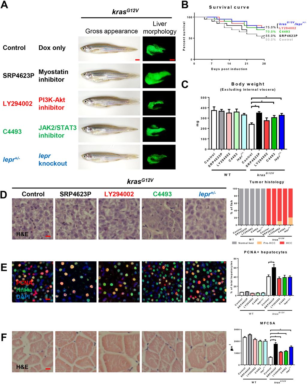
Fig. 6
Effects on hepatocarcinogenesis and muscle wasting of chemical inhibitors targeting downstream of leptin in the signaling pathway. 4-month-old adult male krasG12V zebrafish were treated with dox for 4?weeks with or without SRP4623, LY294002 or C4493 treatment, in comparison with similarly dox-induced krasG12V/lepr+/− fish. In each group, 20 fish were used to initiate the experiments. (A) Gross appearance and liver morphology (left lateral view). Information of targets of chemical inhibitors is shown. (B) Survival curves. (C) Body weight excluding internal viscera. (D) H&E staining of liver sections (left) and quantification of tumor histology (right). (E) IF staining of PCNA (red), Hnf4a (green) and DAPI (blue). Quantification of percentage of proliferating hepatocytes is presented on the right. (F) H&E staining of muscle sections. Quantification of MFCSA is shown on the right. *P<0.05. Scale bars: 2.5?mm in A; 10?μm in D-F.