
|
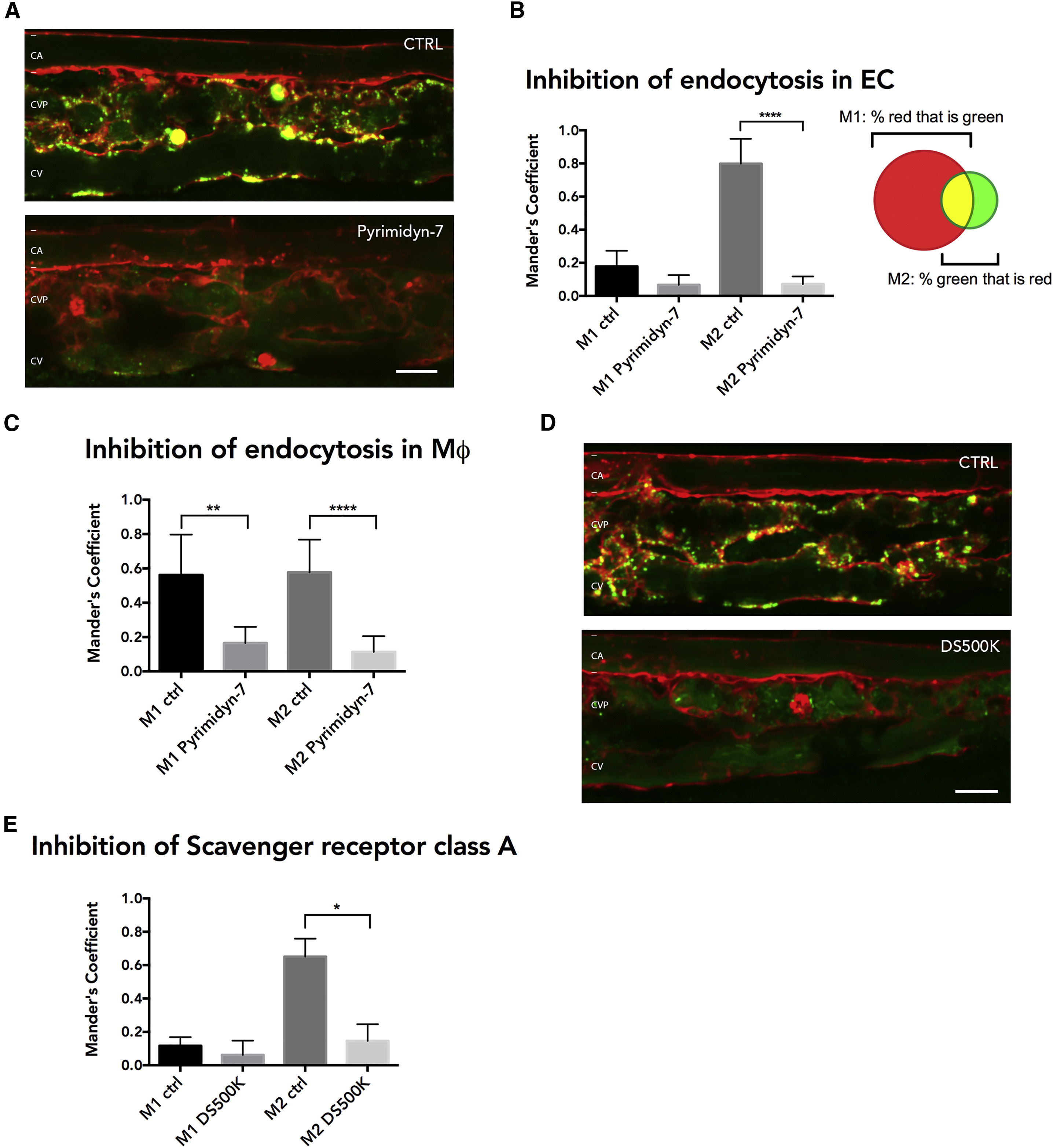
Fig. 6
Characterization of Uptake Mechanism YSL-Exosomes by Endothelial Cells
(A) Close up of the CVP area of 3 dpf Tg(kdrl:Hsa.HRAS-mCherry) zebrafish embryos expressing CD63-pHluorin in the YSL, treated with BafA to show internalized EVs and co-treated with DMSO (CTRL) or dynamin-inhibitor Pyrimidyn-7 (Pyrimidin-7).
(B) Quantification of experiments as performed in (A), showing the overlap coefficient of red with green (M1) and green with red (M2) signal (mean ± SD, ∗∗∗∗p ≤ 0.0001; n ≥ 3; unpaired t test with equal SD).
(C) As in (B), but for macrophages alone (mean ± SD, ∗∗∗∗p ≤ 0.0001; 7 macrophages per condition, n ≥ 3; unpaired t test with equal SD).
(D) Close up of the CVP area of 3 dpf Tg(kdrl:Hsa.HRAS-mCherry) zebrafish embryos expressing CD63-pHluorin in the YSL treated with BafA to show internalized exosomes and non-injected or injected with 500K Dextran Sulfate (DS500K).
(E) Quantification of experiments as performed in (D) (mean ± SD, ∗p ≤ 0.05; n ≥ 2; unpaired t test with equal SD). YSL, yolk syncytial layer; BafA, bafilomycin A; CVP, caudal vein plexus. Scale bars represent 10 μm.
Reprinted from Developmental Cell, 48(4), Verweij, F.J., Revenu, C., Arras, G., Dingli, F., Loew, D., Pegtel, M.D., Follain, G., Allio, G., Goetz, J.G., Zimmermann, P., Herbomel, P., Del Bene, F., Raposo, G., van Niel, G., Live Tracking of Inter-organ Communication by Endogenous Exosomes In Vivo, 573-589.e4, Copyright (2019) with permission from Elsevier. Full text @ Dev. Cell