
|
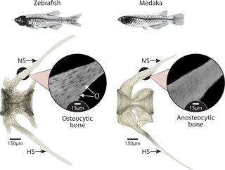
Fig. 1
High-resolution tomography of caudal vertebrae of zebrafish (left) and medaka (right). O, colored in black in the zebrafish scan, show the ubiquity of cells residing in the bone material of zebrafish while being completely absent from medaka bone material. Note that the distal parts of the NS and HS were cropped in the original scans and are only drawn here for reference; therefore, these parts do not contain lacunae in the zebrafish rendering. Inset images show unsegmented tomography slices at a higher magnification. The vertebrae of both species are hourglass shaped along the cranio–caudal axis (see 3D representation in Fig 3A), with NS and HS extending caudally from the vertebral body. HS, hemal spine; NS, neural spine; O, osteocytic lacunae.