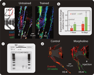
Fig. 6
(a) Double fluorescent ISH, showing SOST (red) and Col1a1 (green) in the spine and adjacent R of untrained (left) and trained (right) medaka (n = 3 for each group). In trained medaka, SOST expression appears down-regulated (as inferred from changes in staining intensity), while osteoblast recruitment is increased. (b) RT-qPCR results of SOST and Col1a1 expression after 1 hour (SOST: swim trained n = 5, untrained n = 7 and Col1a1: swim trained n = 4, untrained n = 6) and 10 days of swim training of medaka (for both SOST and Col1a1: swim trained n = 5, untrained n = 4), (compared to levels in untrained medaka: gray, horizontal “control” line). Results were normalized to the rlp-7 housekeeping gene. Asterisks indicate a significant difference (p < 0.05) between trained and untrained (control) fish. Original raw data can be found Data A. (c) Top: illustration of splice site blocking MO-binding site and primers used to detect morphant transcripts (E1, i1). Bottom: RT-PCR of medaka injected with standard control (C1, C2) and with SOST MO (MO1, MO2). Bands indicative of SOST splice blocking appeared in SOST MO-injected samples (black arrows) and not in the standard control samples. (d) Bone-formation dynamics marked by fluorochromes (red: alizarin red, injected at t = 0 weeks; green: calcein green, injected at t = 2.5 weeks) in vertebra of medaka injected with standard control MO (left) and SOST MO (right). Dashed, vertical white lines mark the border between vertebrae. NSs are cropped in fluorescence images in (d). Col1a1, collagen type I alpha 1; E1, exon-spanning forward primer; HS, hemal spine; ISH, in situ hybridization; i1, intron-spanning reverse primer; MO, vivo-morpholino; NS, neural spine; PCR, polymerase chain reaction; R, radial; RT-PCR, reverse transcription PCR; RT-qPCR, real-time quantitative PCR.

